
Developmental Biology is the study of how a single fertilized egg transforms into a complex multicellular organism through processes like Gametogenesis and Fertilization. This field examines the genetic, molecular, and cellular mechanisms that drive growth, differentiation, and tissue morphogenesis throughout an organism’s life cycle.
Core Principles of Gametogenesis and Cellular Maturation
Gametogenesis refers to the specialized process where diploid germ cells undergo meiosis to produce haploid gametes. In males, this process is spermatogenesis, while in females, it is oogenesis. These biological pathways ensure the continuity of species by preparing cells for successful union.
Spermatogenesis occurs in the seminiferous tubules of the testes. It produces four functional sperm cells from one primary spermatocyte. Oogenesis occurs in the ovaries and results in one functional egg and three non functional polar bodies. This asymmetry ensures the egg retains the cytoplasm and nutrients required for early embryonic life. Understanding these divisions of Developmental Biology is essential for the RPSC Assistant Professor Zoology Paper 1 exam.
As per Developmental Biology, data shows that human spermatogenesis takes approximately 64 to 72 days. During this time, cells transition from spermatogonia to mature spermatozoa. Oogenesis is more prolonged. It begins during fetal development, pauses at Prophase I, and completes only upon Fertilization. These timing differences represent distinct evolutionary strategies for male and female reproductive success.
Molecular Mechanisms of Fertilization
Fertilization is the physical and chemical fusion of male and female gametes to form a zygote. This event triggers the activation of the egg and prevents polyspermy through the cortical reaction. It restores the diploid chromosome number and determines the genetic sex of the individual under the Developmental Biology.
The process involves several specific steps: chemoattraction, acrosome reaction, and membrane fusion. Sperm must penetrate the zona pellucida in mammals or the vitelline envelope in amphibians. Upon contact, the egg releases calcium ions. This calcium wave initiates the completion of meiosis II and starts the metabolic activities of the zygote.
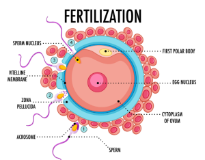
Errors during Fertilization lead to developmental failure or chromosomal abnormalities. Polyspermy occurs if more than one sperm enters the egg. Organisms use fast blocks (electrical changes) and slow blocks (chemical changes) to stop this. Students preparing for RPSC Assistant Professor Zoology syllabus must master these physiological barriers to understand reproductive biology.
Early Embryonic Development and Morphogenetic Movements
Early embryonic development begins with cleavage, a series of rapid mitotic divisions without significant cell growth. This process transforms the zygote into a solid ball of cells called a morula, and later into a hollow ball called a blastula. The pattern of cleavage depends on the amount and distribution of yolk in the egg.
| Cleavage Type | Yolk Distribution | Examples |
| Holoblastic | Isolecithal (Sparse yolk) | Mammals, Echinoderms |
| Meroblastic | Telolecithal (Dense yolk) | Birds, Reptiles |
| Superficial | Centrolecithal (Central yolk) | Insects |
Following blastulation, the embryo undergoes gastrulation. This stage involves morphogenetic movements like invagination, involution, and epiboly. These movements reorganize the single layered blastula into a multilayered structure with three germ layers: ectoderm, mesoderm, and endoderm. Fate maps are used by researchers to track which parts of the early embryo give rise to specific adult tissues.
Organisers and the Process of Organogenesis
Organogenesis is the stage where the three germ layers develop into internal organs. This process relies on induction, where one group of cells influences the development of another. The most famous example is the Spemann-Mangold organiser in amphibians, which induces the formation of the neural tube in Developmental Biology.
The primary organiser secretes signaling molecules like Noggin and Chordin. These proteins inhibit Bone Morphogenetic Proteins (BMPs) to allow ectoderm cells to become neural tissue. Without this specific interaction, the ectoderm would default to skin cells. Organogenesis requires precise spatial and temporal control of gene expression.
You can observe these principles in the development of the vertebrate heart or kidneys. Small changes in signaling gradients can lead to major structural shifts. This section of Developmental Biology highlights the importance of cell communication in building a functional body plan from scratch.
Comparative Development of Frog and Chick
The study of Frog and Chick embryos provides a foundation for vertebrate Developmental Biology. Frogs exhibit external fertilization and an aquatic larval stage known as a tadpole. Chick development occurs inside a shelled egg, requiring specialized membranes to handle waste and respiration.
According to Developmental Biology, Metamorphosis in frogs is a dramatic post embryonic change driven by thyroxine. The tadpole loses its tail and gills while developing lungs and limbs. Chick development focuses on the primitive streak, which serves as the site for gastrulation. Unlike the circular blastopore of frogs, the chick uses this longitudinal groove to internalize cells.
Based on Developmental Biology, chick embryos develop extra embryonic membranes: the amnion, chorion, allantois, and yolk sac. The amnion provides a fluid filled shock absorber. The chorion and allantois work together for gas exchange and calcium transport from the shell. These structures allowed vertebrates to transition from water to land by creating a portable aquatic environment.
Mammalian Placenta Gestation and Parturition
In mammals, the placenta replaces the yolk sac as the primary nutrient source. The placenta is a temporary organ formed from both maternal and fetal tissues. It facilitates the exchange of nutrients, gases, and wastes between the mother and the developing fetus.
Placentas are classified by their shape or the number of tissue layers separating maternal and fetal blood. Humans have a discoid, hemochorial placenta. Gestation is the period from Fertilization to birth. It varies significantly across species. Parturition is the act of giving birth, triggered by a complex hormonal cascade involving oxytocin, cortisol, and prostaglandins.
| Placenta Type | Description | Species Example |
| Diffuse | Villi spread over entire surface | Pig, Horse |
| Cotyledonary | Villi in isolated patches | Cow, Sheep |
| Zonary | Villi in a band around the middle | Dog, Cat |
| Discoid | Villi in a single disc shape | Human, Rodents |
Cell Differentiation and Teratogenesis
Cell differentiation is the process where unspecialized cells become specific cell types like neurons or muscle cells. This is controlled by differential gene expression. Every cell in your body contains the same DNA, but different cells “turn on” different sets of instructions.
Teratogenesis refers to the production of birth defects due to environmental factors. Teratogens include chemicals, radiation, or viruses that disrupt normal development. The timing of exposure is critical. The embryonic period (weeks 3 to 8 in humans) is the most sensitive time because major organ systems are forming.
Common teratogens include alcohol, which causes Fetal Alcohol Syndrome, and thalidomide, which affects limb development. Understanding these disruptions helps scientists identify the “normal” pathways of Developmental Biology. For candidates of RPSC Assistant Professor Zoology Paper 1, this knowledge is vital for clinical and academic applications.
Sex Differentiation in Humans
Sex differentiation is the process of developing the structures that distinguish males from females. In humans, this is initially determined by the presence or absence of the Y chromosome. The SRY gene on the Y chromosome triggers the indifferent gonads to become testes.
If the SRY gene is absent, the gonads develop into ovaries. Hormones then drive secondary sex characteristics in Developmental Biology. Testes produce testosterone and Anti-Mullerian Hormone (AMH). AMH causes the degeneration of female reproductive ducts. Without these hormones, the body naturally follows a female developmental path.
Disruptions in this pathway lead to intersex conditions or disorders of sex development. These cases demonstrate that biological sex is a result of multiple genetic and hormonal steps rather than a single switch. This complexity is a core focus in modern Developmental Biology research.
Mathematical Models and Theorems in Development
Developmental Biology uses mathematical growth models to predict how tissues expand. One common formula is the exponential growth equation for early cleavage:
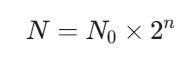
In this equation, N is the final number of cells, N0 is the initial number of cells, and n is the number of division cycles. During the first few hours after Fertilization, embryos follow this rigid mathematical progression.
Another important concept is Wolpert’s French Flag Model. This theorem explains how cells “know” their position based on morphogen concentrations. A cell senses the local concentration of a signal $(C)$ and adopts a fate based on specific thresholds (T1, T2).
If C > T1, the cell becomes Type A.
If T2 < C < T1, the cell becomes Type B.
If C < T2, the cell becomes Type C.
Limitation of the Positional Information Theory
Positional information theory suggests that cells act solely based on their location. This view often fails because it ignores the internal state of the cell. Cells have “memory” based on their previous developmental history. A cell might be in the correct location for a specific signal but cannot respond if it lacks the necessary receptors.
To mitigate this, you must consider competence. Competence is the ability of a cell to respond to an inductive signal. Research shows that timing is just as important as position. If a signal arrives too early or too late, the cell will ignore it. Modern Developmental Biology emphasizes the synergy between external signals and internal genetic readiness.
Conclusion
Mastering Developmental Biology requires a deep understanding of the molecular signals that transform a single cell into a complex organism. From the initial stages of Gametogenesis and Fertilization to the precise structural formation of organs, these biological principles provide the foundation for modern zoological research. VedPrep offers comprehensive resources to help you navigate these complex topics and achieve academic success. By studying these conserved mechanisms, you gain the clarity on key topics such as Developmental Biology needed to excel in the RPSC Assistant Professor Zoology Paper 1 and contribute to the future of biological sciences.
To know more inn detail from our faculty, watch our Youtube Video,
Frequently Asked Questions (FAQs)
What is the role of Gametogenesis in reproduction?
Gametogenesis is the production of haploid sex cells through meiosis. This process ensures that offspring receive the correct number of chromosomes. Spermatogenesis creates sperm in males, while oogenesis produces eggs in females. These specialized cells carry genetic information and cytoplasmic nutrients essential for initiating a new life.
How does Fertilization initiate development?
Fertilization occurs when a sperm and egg fuse to create a diploid zygote. This event triggers metabolic activation and prevents extra sperm from entering the egg. It restores the full set of chromosomes and determines the genetic sex. This union marks the transition from individual gametes to a developing embryo.
What occurs during the cleavage stage of embryonic development?
Cleavage consists of rapid mitotic divisions that occur immediately after fertilization. These divisions increase the number of cells without increasing the overall size of the embryo. This stage transforms the zygote into a morula and eventually a hollow blastula. Cleavage patterns vary across species depending on the yolk distribution.
What is the function of fate maps in embryology?
Fate maps are visual representations showing which parts of an early embryo will become specific adult tissues. Researchers use fluorescent dyes or genetic markers to track cell movements. This tool helps you understand the spatial organization and developmental potential of cells during the blastula and gastrula stages.
How do you classify different types of cleavage?
You classify cleavage based on the amount of yolk present in the egg. Holoblastic cleavage occurs in eggs with little yolk, resulting in complete cell division. Meroblastic cleavage occurs in yolk rich eggs, where division is restricted to a small area. These patterns dictate the physical structure of the early embryo.
How do morphogenetic movements shape the embryo?
Morphogenetic movements occur during gastrulation to rearrange cells into three germ layers. Common movements include invagination, where cells fold inward, and epiboly, where cells spread over the surface. These physical shifts create the ectoderm, mesoderm, and endoderm. This structural reorganization is necessary for organ formation to begin.
What is the significance of the Spemann-Mangold Organiser?
The Spemann-Mangold Organiser is a group of cells that directs the development of neighboring tissues. It produces signaling proteins that inhibit certain growth factors. This interaction induces the formation of the central nervous system. This discovery established the concept of embryonic induction as a fundamental principle of developmental biology.
How does thyroxine regulate metamorphosis in frogs?
Thyroxine is the primary hormone that triggers the transformation from a tadpole to a frog. It stimulates the growth of limbs and the resorption of the tail and gills. The concentration of this hormone determines the timing and success of these physical changes. This process allows the organism to transition from water to land.
Why does polyspermy lead to developmental failure?
Polyspermy happens when multiple sperm fertilize a single egg. This results in an incorrect number of chromosome sets, which disrupts cell division. The embryo cannot coordinate normal growth and usually stops developing early. Eggs use fast electrical blocks and slow chemical blocks to prevent this lethal condition.
What causes disruptions in cell differentiation?
Errors in gene expression or signaling pathways cause differentiation to fail. If a cell cannot receive or process molecular signals, it remains unspecialized or develops into the wrong tissue type. Environmental factors or genetic mutations often cause these disruptions. This failure leads to structural abnormalities or the loss of organ function.
How do teratogens affect different stages of pregnancy?
Teratogens cause birth defects by interfering with specific developmental windows. Exposure during the first trimester is most dangerous because major organ systems are forming. The same chemical might cause limb defects in one week and heart defects in another. Understanding these windows helps you identify the risks of environmental exposure.
What are the specific requirements for RPSC Assistant Professor Zoology Paper 1?
The exam requires detailed knowledge of vertebrate and invertebrate development. You must master topics like placenta types, human sex differentiation, and chick embryology. Focus on the molecular mechanisms of induction and the physiological changes during metamorphosis. Clear understanding of these concepts is essential for scoring well on technical questions.
How does the hemochorial placenta differ from other types?
In a hemochorial placenta, maternal blood comes into direct contact with the fetal chorion. This occurs in humans and rodents. It allows for highly efficient nutrient and gas exchange. Other types, like the epitheliochorial placenta in pigs, have more tissue layers separating the two blood systems.
What is the mathematical relationship in early cleavage cycles?
Cell number increases exponentially during early cleavage. You use the formula N = N0 ×2n to calculate the total cells. N0 is the starting cell count and $n$ is the number of divisions. This predictable growth allows researchers to determine the age and health of an embryo based on cell count.