

Cellular Communication describes the biochemical mechanism by which cells perceive and react to cues from their surroundings or other cells, enabling the coordination of biological activities. This system relies on signaling molecules, specific receptors, and intracellular pathways to regulate vital processes like growth, metabolism, and the regulation of hematopoiesis within complex organisms.
General Principles of Cellular Communication
Cellular Communication lets cells sense and appropriately react to their immediate surroundings. This teamwork forms the foundation for growth, fixing damaged tissue, and defense against disease. Signaling begins when a signaling cell releases a specific ligand. Target cells possess receptors that bind these ligands with high affinity. This binding event triggers a signal transduction pathway.
Four primary types of signaling exist based on the distance the signal travels. Endocrine signaling involves hormones traveling through the bloodstream. Paracrine signaling affects nearby cells. Autocrine signaling occurs when a cell responds to its own secreted factors. Juxtacrine signaling necessitates immediate physical touching of cell surfaces. Each approach guarantees that the RPSC Assistant Professor Zoology Syllabus mandates concerning comprehension of systemic and localized coordination are satisfied.
Regulation of Hematopoiesis through Cellular Signaling
Blood cell generation from stem cells is kept in check through the ongoing activity of hematopoiesis. This procedure primarily takes place within the marrow, subject to precise molecular governance. Cytokines and growth factors serve as the main signaling components. These substances dictate if a stem cell commits to either a myeloid or lymphoid path.
The bone marrow microenvironment furnishes the essential setting for this maturation. Cellular Communication among supporting cells and progenitor cells thwarts the depletion of the precursor supply. Reciprocal circuits guarantee that blood manufacturing aligns with the body’s physiological requirements. For instance, erythropoietin boosts the output of red blood cells when oxygen concentrations fall. This precise governance of blood formation sustains internal balance throughout physical strain or trauma.
Cell Adhesion and Molecular Anchoring
Cell adhesion refers to the linking of a cell to a substrate or an adjacent cell through specialized outer proteins. These connections establish tissue structure and promote physical firmness. Cell adhesion molecules function as the physical links. They belong to four major families: integrins, cadherins, selectins, and the immunoglobulin superfamily.
Cadherins facilitate calcium dependent cell to cell bonding. Selectins mediate the initial sticking of white blood cells to blood vessel walls. These interactions are not static. Cells constantly break and reform these bonds to move or reshape tissues. Strong cell adhesion prevents individual cells from drifting away from their functional units. This stability is crucial for maintaining the integrity of organs and epithelial layers.
The Role of Integrins in Signal Transduction
Integrins are transmembrane receptors that facilitate cell extracellular matrix adhesion. They function as heterodimers consisting of alpha and beta subunits. These proteins serve a dual purpose. They provide physical attachment and act as signaling hubs. Integrins transmit mechanical data from the environment into the cytoplasm.
When integrins bind to the extracellular matrix, they cluster together. This grouping stimulates intracellular enzymes such as Focal Adhesion Kinase. This sequence, termed outside-in signaling, affects cell longevity and growth. Receptors known as Integrins are also involved in inside-out signaling, wherein internal messages alter how strongly the receptor binds to its target molecule. This two-way exchange renders integrins crucial for mending tissues and movement of immune system cells.
Functions of the Extracellular Matrix
The extracellular matrix material is composed of a complex web of biomolecules, both protein and carbohydrate, released by cells. This structure furnishes physical underpinning for tissues and impacts cellular actions. Key elements feature collagen, elastin, fibronectin, and laminin. Every constituent bestows distinct physical characteristics. Collagen imparts resistance to stretching, whereas elastin permits tissues to spring back.
It functions as a storage pool for growth factors. Cells detect the rigidity of the surrounding matrix via their receptors. This mechanical signaling dictates whether a cell will proliferate or stay dormant in Cellular Communication. Within the RPSC Assistant Professor Zoology Syllabus, the extracellular matrix is emphasized as a vital controller of tissue formation. Alterations in this matrix composition frequently result in disease states such as scarring or the spread of cancer.
Gap Junctions and Direct Cytoplasmic Contact
Direct routes for Cellular Communication are furnished by gap junctions. These conduits permit the transit of charged particles, sweeteners, and minor message substances such as cyclic AMP. Every gap junction is composed of a pair of half-channels known as connexons. Six connexin proteins form one connexon. When two connexons from neighboring cells align, they create a continuous aqueous pore.
This direct connection synchronizes the electrical activity of cell clusters in Cellular Communication. In cardiac muscle, gap junctions ensure that the entire heart contracts as a single unit. They also play a role in metabolic coupling. A cell with low nutrient levels can receive glucose from a healthy neighbor through these channels. This shared resource system enhances the resilience of the tissue.
Neurotransmission and its Regulation
Nerve signal transmission is a unique type of cell-to-cell talk in the nervous structure. This process features the swift dispatch of chemical signals named neurotransmitters across a synaptic gap in Cellular Communication. Once an electrical impulse arrives at the axon ending, it prompts the entry of calcium particles. This entry leads to synaptic sacs merging with the membrane at the front of the synapse.
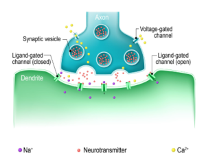
The freed signaling molecules attach to acceptance sites on the receiving nerve cell. This connection triggers an electrical or chemical reaction. Modulating takes place via a number of methods. Recycling mechanisms pull back neurotransmitters from the gap between cells. Certain catalysts also break down the compounds to end the message. This exact management stops excessive exciting of nerve cells. The RPSC Assistant Professor Zoology Syllabus emphasizes these regulatory steps as fundamental to nervous system function in Cellular Communication.
Critical Perspectives on Signaling Redundancy
Numerous biological frameworks posit that one ligand yields one foretellable result. Yet, in the realm of Cellular Communication, data point towards signal transduction routes possessing substantial overlap. Should one route be inhibited, the cell frequently initiates a corresponding track to attain the identical endpoint. This duplication complicates efforts to focus on particular ailments.
Blocking just one receptor seldom halts a biological process entirely. This constraint accounts for why certain drug therapies lose efficacy eventually. To overcome this, current approaches emphasize dampening multiple points. Recognizing that Cellular Communication operates as a network instead of a stepwise sequence is crucial for successful intervention.
Cellular Communication Data and Theoretical Models
Mathematical models help predict how cells respond to varying concentrations of signals. The following table outlines key concepts used to quantify these interactions.
| Concept Name | Mathematical Expression | Application |
| Scatchard Equation | $r/C = nK – rK$ | Determines receptor binding affinity and number of sites |
| Hill Equation | $\theta = \frac{[L]^n}{K_d + [L]^n}$ | Describes the degree of cooperativity in ligand binding |
| Fick’s Law of Diffusion | $J = -D \frac{dc}{dx}$ | Models the movement of signaling molecules through the extracellular matrix |
| Nernst Equation | $E = \frac{RT}{zF} \ln \frac{[ion]_{out}}{[ion]_{in}}$ | Calculates the equilibrium potential during neurotransmission |
Practical Scenario: Bone Marrow Transplant and Hematopoiesis
A bone marrow transplant provides a real world example of Cellular Communication in action. When a patient receives donor stem cells, these cells must find their way to the bone marrow. This process is called homing. The donor cells use integrins to stick to the blood vessel walls near the marrow.
Once inside the marrow, the new cells interact with the extracellular matrix. These corporeal interactions initiate the control of blood cell formation. The donated cells start to multiply and generate fresh blood components according to the chemical prompts they get from the recipient’s system. Should the molecules responsible for cell attachment not operate properly, the graft will not take hold as the cells lack the ability to secure themselves. The positive outcome relies wholly on the molecular exchange between the donor cells and the surrounding host milieu.
Conclusion
Exploring Cellular Communication uncovers the intricate molecular systems that maintain living organisms via accurate signaling and direct contacts. Gaining proficiency in controlling blood cell formation and the workings of the tissue surrounding cells offers a core benefit for applicants studying the RPSC Assistant Professor Zoology curriculum. VedPrep provides comprehensive academic resources to help students navigate these complex cellular pathways effectively. Understanding how integrins and gap junctions maintain tissue integrity ensures a complete perspective on modern cell biology. By analyzing these systems, you gain the insight necessary to interpret advanced physiological processes in both research and clinical settings.
To learn more from our faculty, watch our Youtube video:
Frequently Asked Questions (FAQs)
How does paracrine signaling differ from endocrine signaling?
Paracrine signaling targets cells located in the immediate vicinity of the signaling cell. In contrast, endocrine signaling releases hormones into the bloodstream to reach distant organs. Paracrine actions are fast and local, while endocrine actions are systemic and slower due to blood circulation times.
What roles do gap junctions play in cell to cell contact?
Gap junctions create direct physical channels between the cytoplasm of adjacent cells. These pores allow ions and small signaling molecules to pass freely. This connectivity enables groups of cells to synchronize their electrical activity, which is vital for the rhythmic contraction of heart muscle.
What defines the extracellular matrix in biological tissues?
The extracellular matrix is a non cellular network of proteins and polysaccharides. It provides a physical scaffold for cells and stores growth factors. This matrix influences cell behavior by communicating mechanical stiffness and chemical signals to the internal cytoskeleton through specialized surface receptors.
How do cells identify specific signals in a crowded environment?
Cells express specific proteins called receptors that bind to complementary ligands with high affinity. This binding specificity ensures that only the intended target cells respond to a particular signal. Even if multiple signals are present, a cell only reacts if it possesses the corresponding receptor.
How is the regulation of hematopoiesis managed in the bone marrow?
The regulation of hematopoiesis relies on a specialized microenvironment known as the niche. Stromal cells in the bone marrow release cytokines that signal hematopoietic stem cells to either remain dormant or divide. This ensures a steady supply of blood cells based on current body needs.
How do integrins facilitate cell adhesion to the extracellular matrix?
Integrins act as transmembrane bridges connecting the extracellular matrix to the internal cytoskeleton. They bind to proteins like fibronectin and laminin outside the cell. Once bound, they cluster and initiate intracellular signals that stabilize the cell's position and govern its movement or survival.
What mechanisms regulate neurotransmission at the synapse?
Regulation occurs through the precise control of neurotransmitter release and removal. Once a neurotransmitter binds to a postsynaptic receptor, the signal is terminated by reuptake into the presynaptic neuron or enzymatic degradation. This prevents continuous signaling and allows the nervous system to process discrete pieces of information.
How does cell adhesion affect the movement of immune cells?
Immune cells use dynamic cell adhesion to exit blood vessels and enter infected tissues. They use selectins to slow down and integrins to form firm attachments to the vessel wall. This controlled sticking and unsticking allows the cells to migrate precisely toward the site of inflammation.
What causes failures in the regulation of hematopoiesis?
Disruptions often stem from mutations in cytokine receptors or defects in the bone marrow niche. If the signaling between stromal cells and stem cells breaks down, it can lead to bone marrow failure or leukemia. In these cases, the balance between cell self renewal and differentiation is lost.
Why might a cell fail to respond to a hormone signal?
A cell will not respond if it lacks the specific receptor for that hormone. Downregulation of receptors also reduces sensitivity. If the internal signal transduction proteins are mutated or inhibited, the message stops at the membrane and fails to produce a physiological response.
What are the consequences of abnormal extracellular matrix stiffness?
Excessive stiffness in the extracellular matrix often signals fibrosis or cancer progression. Cells sense this mechanical change and may begin to divide uncontrollably or migrate inappropriately. This mechanical feedback loop can override normal chemical signals that usually limit cell growth.
What is the significance of signal redundancy in Cellular Communication?
Redundancy involves multiple signaling pathways that achieve the same biological outcome. This provides a backup system if one pathway is damaged. In medical treatments, redundancy often leads to drug resistance because the cell bypasses the inhibited route to continue its functions.
How do different adhesion molecules cooperate during tissue formation?
Tissue formation requires the coordinated effort of cadherins for cell to cell binding and integrins for matrix attachment. These molecules work in sequence to organize cells into functional layers. For example, during embryonic development, switching between different adhesion types allows cells to move and then settle.
What is the role of cyclic AMP in gap junction communication?
Cyclic AMP acts as a secondary messenger that can pass through gap junctions. This allows a signal generated in one cell to trigger a response in its neighbors. This shared signaling helps large groups of cells respond as a single functional unit to a stimulus.