

The ultrastructure of bacteria describes the fine internal and surface arrangement of bacterial cells, visible through electron microscopes. This encompasses the cell boundary, internal fluid, genetic material area, and external features such as flagella. Knowing these elements is crucial for the RPSC Assistant Professor Botany Syllabus because it underpins how microbes are grouped and how they cause disease.
Exploring the Ultrastructure of Bacteria is a fundamental aspect of microbiology and a crucial part of both paper I and paper II in the RPSC Assistant Professor Botany curriculum. Moving past the basic depictions often seen in entry-level biology, the inner and outer organization of a bacterial cell demonstrates a complex, structured system equipped for thriving in harsh conditions. This in-depth look utilizes tools beyond the light microscope to investigate the molecular makeup of the cell’s outer covering, the internal cytoplasm, and specific hereditary elements. Scrutinizing these elements allows for comprehension of how prokaryotes sustain efficient metabolism without the organelle compartments characteristic of eukaryotic cells.
Understanding the Ultrastructure of Bacteria is crucial for pinpointing disease-causing agents and creating successful remedies for crop ailments. Deviations like L-form bacteria and Mycoplasma demonstrate the remarkable adaptability of bacterial shape. These organisms lacking a cell wall complicate standard categorization and demand tailored methods for their detection. Whether you are examining the 70S protein synthesizers or the twisted genetic material in the nucleoid region, every component of the architecture yields insight into how these extraordinarily resilient life forms thrive. VedPrep supplies the expert materials and thoughtful approach necessary to conquer these intricate phytopathology ideas for career and scholarly achievement.
Core Components of the Bacterial Cell Envelope
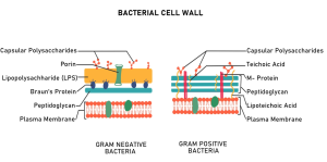
Regarding Ultrastructure of Bacteria, the envelope surrounding the bacterial cell is an intricate, multi-tiered assembly safeguarding the microbe against external pressures. This envelope comprises the inner cytoplasmic membrane, the cell wall, and in certain types, an external capsule. The makeup of the cell wall dictates the cell’s Gram staining outcome. Species classified as Gram-positive feature a substantial peptidoglycan stratum, whereas Gram-negative bacteria exhibit a less substantial layer alongside an external membrane incorporating lipopolysaccharides.
The cytoplasmic membrane acts as a selective barrier in Ultrastructure of Bacteria. It regulates the transport of nutrients and waste products. Bacterial membranes, distinct from those in eukaryotes, typically do not contain sterols such as cholesterol, with the exception of Mycoplasma. The membrane also contains the machinery for DNA duplication and energy generation, as bacteria do not possess mitochondria. This streamlined structure enables bacteria to sustain rapid metabolic activity across diverse settings.
Essential Internal Structures and the Nucleoid
Based on ultrastructure of bacteria, Bacteria lack a membrane bound nucleus, which defines their prokaryotic nature. The hereditary substance is present as one, ring-like, two-stranded DNA entity situated in a poorly defined area termed the nucleoid. This arrangement is a main point of emphasis in the RPSC Assistant Professor Botany Syllabus for both Paper 1 and Paper 2. The DNA is extensively coiled and bonded with proteins other than histones so it can be contained within the limited cellular space.
As per Ultrastructure of Bacteria, the internal fluid, or cytoplasm, holds ribosomes, inclusion bodies, and plasmids. Bacterial ribosomes are of the 70S variety, made up of 50S and 30S components. These are smaller than the 80S ribosomes seen in eukaryotic cells. Plasmids are accessory strands of DNA that frequently bear genes providing resistance to antibiotics. Inclusion bodies serve as storage vessels for nutrients like glycogen, lipids, or phosphate. These internal features ensure survival during periods of nutrient limitation.
Characteristics of L-form Bacteria and Cell Wall Deficiencies
L-form bacteria are specialized variants that have lost their ability to produce a cell wall. These structures emerge naturally or via prompting by antimicrobials such as penicillin, which interfere with cell envelope creation. In contrast to standard bacteria, L-variants display varied shapes as they do not possess the firm structure given by peptidoglycan. They can endure in salt concentrations that usually lead to bursting in cells possessing a wall.
L-form bacteria pose a considerable hurdle in clinical environments concerning bacterial ultrastructure. They frequently evade typical antibiotic therapies since the essential target, the cell wall, is absent. These microbes are capable of returning to their typical walled form once the stressing agent is withdrawn. Studying L-form bacteria offers understandings into bacterial development and the fundamental necessities for cell viability. Their study is a distinct requirement for candidates preparing for the RPSC Assistant Professor Botany Syllabus.
Mycoplasma and the Smallest Living Prokaryotes
Mycoplasma species are the smallest known free living microorganisms in ultrastructure of bacteria. They are unique among bacteria because they naturally lack a cell wall. This lack of a cell wall makes them naturally resilient to antibiotics such as beta-lactams. Their outer membrane includes sterols, a characteristic typically seen in eukaryotes, which supplies needed balance against osmotic pressure. Mycoplasma are significant pathogens in both humans and plants.
Regarding plant diseases, organisms resembling mycoplasma induce conditions like brinjal little leaf and witches broom. Based on their ultrastructure of bacteria, transmission usually occurs via insect carriers such as leafhoppers. Due to their minute dimensions, they can traverse filters that retain other bacteria. Grasping the nature of Mycoplasma is crucial for excelling in the microbiology segment of the RPSC Assistant Professor Botany Curriculum. Their simplified ultrastructure serves as a model for synthetic biology.
Surface Appendages and Motility Mechanisms
Bacteria use specialized surface structures for movement and attachment in ultrastructure of bacteria. Flagella are elongated, slender filaments made up of the protein flagellin. They spin like a propeller, propelling the cell through fluid environments. The pattern of flagella, such as monotrichous, lophotrichous, or peritrichous, serves as a distinguishing characteristic employed in bacterial identification.
Pili and fimbriae are brief, filament-like formations. Fimbriae aid microbes in fastening to substrates or host cells, a vital stage in bacterial infection at the ultrastructure of bacteria. Sex pili are key to conjugation, the mechanism for transferring genetic material between bacterial populations. These extensions allow bacteria to perceive and react to their physical surroundings, thus promoting their persistence in challenging ecological settings.
Summary of Bacterial Ultrastructure for Competitive Exams
The following table summarizes the key structural components and their functions relevant to the RPSC Assistant Professor Botany Syllabus.
| Structure | Composition | Primary Function |
| Cell Wall | Peptidoglycan (Murein) | Provides shape and osmotic protection |
| Plasma Membrane | Phospholipid bilayer | Transport and ATP synthesis |
| Nucleoid | Circular DNA | Genetic information storage |
| Ribosomes | 70S (30S + 50S) | Protein synthesis |
| Flagella | Flagellin protein | Locomotion and chemotaxis |
| Capsule | Polysaccharides | Protection from phagocytosis |
| Plasmids | Extra chromosomal DNA | Antibiotic resistance and conjugation |
Conclusion
Grasping the ultrastructure of bacteria is a core necessity for anyone preparing for the RPSC Assistant Professor Botany curriculum. This understanding offers the basic blueprint for comprehending how these prokaryotic life forms operate, multiply, and relate to their surroundings. Ranging from the firm peptidoglycan coverings of typical bacteria to the flexible shapes of L-form varieties and Mycoplasma, every structural shift signifies an evolutionary adjustment to particular habitats or dangers to survival. Concentrating on these cellular specifics permits the capability to distinguish between intricate disease agents and grasp the processes behind plant ailments.
Learning environments such as VedPrep furnish the organized direction and superior study materials needed to confidently tackle these complex areas of botany. Understanding the subtleties of the microbial cell coating, the arrangement of the 70S ribosome, and the singular absence of a cell wall in Mycoplasma guarantees a thorough comprehension of the demands for both Paper 1 and Paper 2. As you continue your preparation, use these ultrastructure of bacteria insights to bridge the gap between theoretical microbiology and practical applications in plant pathology and biotechnology. A deep dive into these microscopic details is the most effective way to secure a competitive edge in professional academic examinations.
To know more in details from our expert team, watch our Youtube video:
Frequently Asked Questions (FAQs)
How does the bacterial cell wall function?
The cell wall provides structural integrity and prevents osmotic lysis. In Gram positive species, a thick layer of peptidoglycan maintains shape. Gram negative bacteria possess a thinner layer and an outer membrane containing lipopolysaccharides. This barrier regulates the entry of toxic substances and determines the bacterial response to staining.
What are the primary characteristics of Mycoplasma?
Mycoplasma species are the smallest free living organisms and lack a cell wall entirely. Their plasma membrane contains sterols for stability, which is rare in prokaryotes. This lack of a wall makes them resistant to penicillin and other beta lactam antibiotics. They cause significant diseases in both animals and plants.
What is the nucleoid in bacterial cells?
The nucleoid is an irregularly shaped region containing the primary genetic material. Unlike eukaryotes, bacteria do not have a nuclear membrane. The DNA is typically a single, circular, double stranded molecule. It remains compact through supercoiling and interactions with nucleoid associated proteins to fit within the small cellular volume.
How do you identify L-form bacteria?
You identify L-form bacteria by their pleomorphic shape and lack of a cell wall under specific growth conditions. These forms arise when walled bacteria lose their peptidoglycan layer due to antibiotic stress or enzyme action. They require osmotically stable media to prevent bursting. Detection often involves specialized culturing techniques and electron microscopy.
How does bacterial ultrastructure influence antibiotic selection?
Antibiotic selection depends on the presence of specific targets within the bacterial ultrastructure. Penicillin targets peptidoglycan synthesis, making it effective against walled bacteria but useless against Mycoplasma. Aminoglycosides target the 30S subunit of 70S ribosomes. Understanding the target site ensures you choose an antibiotic that effectively disrupts the pathogen.
What is the significance of plasmids in microbiology?
Plasmids are extra chromosomal DNA loops that provide selective advantages like antibiotic resistance or toxin production. You find them frequently in Gram negative bacteria. They replicate independently of the chromosomal DNA. Scientists use plasmids as vectors in genetic engineering to introduce new genes into bacterial hosts for industrial or medical purposes.
How do inclusion bodies aid bacterial survival?
Inclusion bodies store essential nutrients as insoluble granules within the cytoplasm. Bacteria accumulate substances like glycogen, poly beta hydroxybutyrate, or sulfur when resources are abundant. During periods of starvation, the cell mobilizes these reserves to maintain metabolism. This storage mechanism allows bacteria to survive in fluctuating and nutrient poor environments.
How does the RPSC Assistant Professor Botany Syllabus categorize bacteria?
The RPSC syllabus groups bacteria based on ultrastructure, reproduction, and pathological roles. It emphasizes the differences between typical walled bacteria, L-form bacteria, and wall less Mycoplasma. Candidates must understand these structural distinctions to explain plant diseases and microbial ecology. The syllabus connects these physical traits to broader biological functions.
Why do Gram negative bacteria resist certain detergents?
The outer membrane of Gram negative bacteria acts as a selective permeability barrier. Lipopolysaccharides and porin proteins restrict the passage of large or hydrophobic molecules like detergents and certain antibiotics. This structural defense mechanism protects the underlying cell wall and plasma membrane from chemical damage in hostile environments like the human gut.
Why does Mycoplasma cause chronic infections?
Mycoplasma causes chronic infections because its lack of a cell wall allows it to mimic host cell membranes. This structural similarity helps the pathogen evade the host immune system. Additionally, their small size and flexibility allow them to sequester in tight spaces within tissues where immune cells or standard concentrations of antibiotics struggle to reach.
How do L-form bacteria survive penicillin treatment?
L-form bacteria survive penicillin because they lack the peptidoglycan target that the drug attacks. While the drug destroys the walled version of the bacteria, L-forms persist in a dormant or slow growing state. Once the antibiotic pressure is removed, many L-forms can resynthesize their cell walls and cause a relapse of the infection.
How do magnetosomes function in specific bacteria?
Magnetosomes are specialized inclusion bodies containing magnetite or greigite crystals. These structures allow bacteria to sense the magnetic field of the Earth, a process called magnetotaxis. By aligning themselves with magnetic lines, these bacteria can move vertically to find optimal oxygen concentrations in aquatic sediments. This demonstrates advanced structural specialization in prokaryotes.
What is the role of the S-layer in bacterial envelopes?
The S-layer is a paracrystalline surface layer composed of identical proteins or glycoproteins. It acts as a molecular sieve, protecting the cell from predatory bacteria or bacteriophages. It also provides structural rigidity in some species. The S-layer represents a highly organized protein assembly that is often lost in laboratory cultures but critical in nature.
How do endospores differ from vegetative cells?
Endospores are highly resistant, dormant structures formed within some Gram positive bacteria. Their ultrastructure includes a thick cortex and a proteinaceous spore coat that protects the core DNA. They exhibit almost no metabolic activity and can survive extreme heat, radiation, and desiccation for centuries. This makes them a primary concern in food safety and sterilization.