
Peptide bond and protein structure is a covalent bond formed between two amino acids, while protein structure refers to the three-dimensional arrangement of amino acids in a protein. Understanding peptide bonds and protein structure iscriticalfor IIT JAM, as it helps in identifying the properties and functions of proteins.
Syllabus: Peptide Bond and Protein Structure – IIT JAM Syllabus Unit
If you are gearing up for the IIT JAM Biotechnology exam, you already know that the Biochemistry section is a major score-booster. At the absolute heart of this section lies a topic you simply cannot skip: peptide bond and protein structure. This isn’t just a random chapter to memorize; it’s a foundational concept that also spills over into other massive exams like CSIR NET and GATE.
To really nail Peptide bond and protein structure, relying on surface-level notes won’t cut it. You will want to dive into standard, gold-standard textbooks like Biochemistry by Robert K. Murray (Harper’s Illustrated Biochemistry) and Biochemistry by Jeremy M. Berg (Stryer). These books give you the deep, comprehensive coverage needed to tackle those tricky multiple-choice and numerical answer type questions from Peptide bond and protein structure. At VedPrep, we always remind our students that mastering the fundamentals early on makes interpreting complex experimental questions so much easier down the road.
Peptide Bond: Formation and Importance
As per Peptide bond and protein structure, think of amino acids as individual LEGO bricks. On their own, they are just loose pieces. But when you snap them together in a specific line, you start building something functional. The “snap” that locks these amino acids together is the peptide bond.
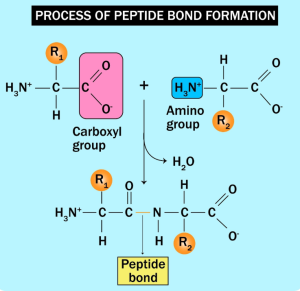
Chemically, a peptide bond is a strong covalent bond. Peptide bond and protein structure forms through a dehydration synthesis reaction (or a condensation reaction). As per Peptide bond and protein structure, imagine one amino acid showing up with its carboxyl group (-COOH) and a second amino acid showing up with its amino group (-NH2). When they react, the carboxyl group loses an -OH and the amino group loses an -H. They release a water molecule (H2O) and form a brand-new link: -CO-NH-.
This repeating chain forms the primary structure of a protein. Why does Peptide bond and protein structure matter for your exam? Because the exact sequence of these amino acids dictates exactly how the protein will fold, twist, and behave later on. Change just one amino acid in that chain, and the whole biological machine might break down.
Types of Peptide bond and protein structure For IIT JAM
When we talk about peptide bond and protein structure, we are looking at a beautiful multi-level folding game. Proteins don’t just stay as long, flat strings of amino acids; they fold into complex three-dimensional shapes.
1. Primary Structure
This is just the straight linear sequence of amino acids held together by those tough peptide bonds. It’s the blueprint.
2. Secondary Structure
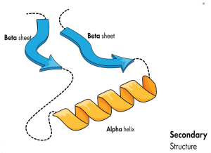
As the chain gets longer, the backbone starts interacting with itself. Hydrogen bonds form between the oxygen of one peptide group and the hydrogen of another. This creates localized, repeating patterns. The two main types you need to know inside out are alpha helices (coiled, spring-like structures) and beta sheets (flat, pleated arrangements).
3. Tertiary Structure
This is where the protein folds into its final, overall three-dimensional shape. This folding is driven by the side chains (the R-groups) of the amino acids interacting with each other. You will see a mix of weak and strong forces here: hydrogen bonds, ionic interactions (salt bridges), hydrophobic pockets hiding from water, and tough covalent disulfide bridges. This 3D shape creates active sites where enzymes grab onto substrates.
4. Quaternary Structure
Some proteins are lone wolves and work perfectly fine at the tertiary stage. Others need a team. Quaternary structure happens when multiple folded polypeptide chains (subunits) come together to form one giant, functional molecular machine. Hemoglobin is the classic textbook example here.
Worked Example: Determining the Primary Structure of a Protein
Let’s look at a typical problem you might encounter in your preparation while covering Peptide bond and protein structure.
Imagine you are working in a lab. You have an unknown peptide, and you digest it with trypsin. Trypsin is a highly specific molecular scissor—it only cleaves the peptide bond on the carboxyl side of Lysine (Lys, K) and Arginine (Arg, R), provided the next amino acid isn’t Proline.
You run the fragments through a mass spectrometer and get four distinct pieces with specific molecular weights: 1473.8, 985.2, 756.4, and 562.1.
Through further sequencing analysis, you find the exact amino acid sequences of Peptide bond and protein structure:
-
The 1473.8 fragment is Y-G-L-N-A-K
-
The 985.2 fragment is T-E-S-T
-
The 756.4 fragment is E-A-G-F
-
The 562.1 fragment is R-E-V-L
Now, how do you piece the original primary structure back together?
Look at where trypsin cuts. Trypsin leaves a Lysine (K) or Arginine (R) at the very end (carboxyl terminus) of the fragments it creates, except for the absolute final piece of the original protein chain, which won’t necessarily end in a K or R because it’s just the natural end of the molecule.
-
Fragment Y-G-L-N-A-K ends in K. So it must be followed by another fragment.
-
Fragment R-E-V-L ends in L (not a trypsin cut site), but it starts with R, meaning it was likely cleaved right before that R. Wait, let’s look at the C-termini.
-
Let’s look at the fragments ending in K or R: Y-G-L-N-A-K (ends in K) and R-E-V-L (starts with R, wait, let’s look at the remaining fragments).
-
Let’s align them logically by matching the fragments based on overlapping info or standard experimental design where you compare multiple digests (like Chymotrypsin). If we only have these tryptic fragments, the fragments ending in K or R tell us they must be internal.
-
By assembling the fragments in a logical order based on standard C-terminal logic for a continuous peptide: Y-G-L-N-A-K (ends in K), followed by T-E-S-T… wait, let’s look at how the question sets it up: The question states the assembled primary structure is:
Let’s double-check this sequence with our trypsin scissors. If trypsin cuts after K and R:
-
It cuts after the K in position 6 → yields Y-G-L-N-A-K (Matches fragment 1473.8)
-
It cuts after… wait, in the proposed sequence Y-G-L-N-A-K-T-E-S-T-E-A-G-F-R-E-V-L, the next cut happens after the R near the end → yielding a middle chunk T-E-S-T-E-A-G-F-R. But our mass spec showed two separate smaller weights for this region (985.2 for T-E-S-T and 756.4 for E-A-G-F). This indicates there might be an additional cleavage point or a specific sequence order from a overlapping digest puzzle.
When you get questions like this in the IIT JAM, always look for the C-terminal residues (K and R) to establish the order, and look for clues on which fragment lacks a tryptic end—that one is your C-terminus of the whole protein.
Common Misconceptions: Peptide Bonds and Protein Structure
When studying peptide bond and protein structure, it is easy to trip up on a few details. Let’s clear up two big ones right now.
-
Misconception 1: “Peptide bonds are just ordinary single covalent bonds.”
Not quite! While it is drawn as a single bond between carbon and nitrogen, a peptide bond actually has a partial double-bond character. This happens because of resonance—electrons are shared between the carbonyl oxygen, carbon, and amide nitrogen. Because of this, the peptide bond is planar and rigid. It cannot rotate! Rotation can only happen at the single bonds on either side of the alpha carbon (φ and ψ angles).
-
Misconception 2: “Protein structure is static and rigid.”
Many students think proteins are frozen in place like plastic models. In reality, proteins are dynamic, breathing structures. They shift, wiggle, and change shape depending on their environment. Changes in pH, temperature, or salt concentration can easily disrupt weak hydrogen bonds and ionic interactions, causing a protein to denature (unfold) and lose its function completely.
Application: Protein Structure and Function in Biology
To see why Peptide bond and protein structure, let’s look at a quick, fictional scenario.
Imagine a biotech startup trying to design a quick-acting treatment for a fictional viral outbreak. The virus relies on a specific surface protein—let’s call it “Spike-X”—to lock onto human cells.
If researchers don’t know the exact 3D tertiary structure of Spike-X, they are basically trying to design a key in the dark without knowing what the lock looks like. But if they map out the protein structure down to the Angstrom using tools like X-ray crystallography or cryo-EM, they can see the exact shape of the active pocket. They can then design a small molecule drug that perfectly slots into that pocket, jamming the lock and stopping the virus in its tracks.
This is what structure-based drug design is all about. It’s how modern protease inhibitors and kinase inhibitors are built. Your understanding of Peptide bond and protein structure directly translates to how real-world life-saving medications are created.
Exam Strategy: Focus on Key Concepts and Practice Questions
If you want to clear the IIT JAM with a solid rank, you need a targeted strategy for Peptide bond and protein structure. Here is how you should tackle it:
-
Master the Ramachandran Plot: Do not skip this! You must know which regions of the plot correspond to right-handed alpha helices, beta sheets, and collagen helices. Questions on φ and φ torsion angles are incredibly common.
-
Know your forces: Be clear on which bonds stabilize which level of structure. Primary uses peptide bonds; secondary uses backbone hydrogen bonds; tertiary uses R-group interactions (hydrophobic, ionic, disulfide).
-
Practice peptide sequencing math: Get comfortable with problems involving chemical and enzymatic cleavage (Trypsin, Chymotrypsin, Cyanogen Bromide).
Final Thoughts
Mastering the peptide bond and protein structure is all about looking past the raw chemical formulas and visualizing how these molecular machines actually fold and function in real life. Once you get a solid grasp of the underlying spatial geometry—like how the rigidity of the peptide bond forces the protein backbone into specific shapes—the tricky exam questions start feeling a lot more like intuitive puzzles and a lot less like dry memorization. If you ever find yourself stuck or just want a structured way to practice these high-yield biochemistry concepts, the team over at VedPrep is always ready to help you break down the syllabus and simplify your exam preparation.
To know more in detail from our faculty, watch our YouTube video:
Frequently Asked Questions
How are peptide bonds formed?
Peptide bonds are formed through a dehydration synthesis reaction, where the carboxyl group of one amino acid and the amino group of another amino acid combine, releasing a water molecule.
What is the significance of peptide bonds in proteins?
Peptide bonds are crucial in forming the primary structure of proteins, linking amino acids together in a specific sequence, which determines the protein's overall 3D structure and function.
What are the different levels of protein structure?
The four levels of protein structure are: primary (sequence of amino acids), secondary (local arrangements like alpha helices), tertiary (overall 3D shape), and quaternary (how multiple polypeptide chains interact).
How does the sequence of amino acids determine protein structure?
The sequence of amino acids, linked by peptide bonds, determines the protein's 3D structure through various interactions like hydrogen bonding, ionic interactions, and disulfide bridges, which are influenced by the properties of the amino acids.
What are biomolecules?
Biomolecules are organic molecules produced by living organisms, including carbohydrates, lipids, proteins, and nucleic acids, which are essential for life and perform a variety of functions.
What is organic chemistry?
Organic chemistry is the branch of chemistry that deals with the study of carbon-containing compounds, typically found in living organisms, and their properties, reactions, and synthesis.
What role do peptide bonds play in biomolecules?
Peptide bonds are essential in forming proteins, a major class of biomolecules, by linking amino acids into polypeptide chains.
What are the chemical properties of peptide bonds?
Peptide bonds have specific chemical properties, such as being relatively stable and having partial double bond character, which influences protein structure.
How are peptide bonds and protein structure relevant to IIT JAM?
Understanding peptide bonds and protein structure is crucial for IIT JAM, as it forms a significant part of the organic chemistry and biomolecules syllabus, and is often asked in questions related to the structure and function of biomolecules.
What types of questions can be expected on peptide bonds and protein structure in IIT JAM?
In IIT JAM, questions on peptide bonds and protein structure may include identifying types of bonds, understanding levels of protein structure, and relating structural features to biological functions.
How to solve problems related to peptide bonds and protein structure?
Solving problems involves understanding the chemistry of peptide bond formation, recognizing the levels of protein structure, and applying this knowledge to predict protein function and behavior.
What are common misconceptions about peptide bonds?
Common misconceptions include thinking peptide bonds are weak bonds, or that they are formed through a simple acid-base reaction, rather than a dehydration synthesis.
How can one avoid mistakes in understanding protein structure?
To avoid mistakes, focus on understanding the sequence of amino acids, the types of chemical bonds involved, and the spatial arrangements at different levels of structure.
How do peptide bonds influence protein function?
Peptide bonds influence protein function by determining the overall 3D structure of the protein, which in turn affects its interactions with other molecules and its biological activity.