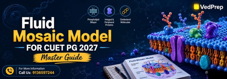

Fluid Mosaic Model for CUET PG Zoology 2027
This Fluid Mosaic model for CUET PG Zoology 2027 describes the structure and behaviour of the plasma membrane. Singer and Nicolson (1972) proposed the model, which describes membranes as being made up of a fluid phospholipid bilayer with proteins embedded in it that are free to move around. This topic is particularly important for CUET PG Zoology because of the connection between membrane transport, cell signaling, organelle function and molecular biology.
Importance of the Fluid Mosaic Model for CUET PG Zoology 2027
The fluid-mosaic model is the conceptual underpinning of membrane biology and is frequently asked in the zoology entrance exams. Questions often combine membrane transport, communication between cells, osmosis, diffusion, endocytosis and membrane proteins. With a comprehensive comprehension of the model, students may tackle both factual and analytical questions in CUET PG Zoology.
The plasma membrane regulates the flow of materials in and out of cells. The shape of the membrane impacts how cells recognise signals, maintain homeostasis, and communicate with neighbouring tissues. These functions make the fluid-mosaic model no longer a topic in isolation. The idea is explicitly tied to physiology, immunology, molecular genetics, and biochemistry.
Many students know the parts of membranes, but they don’t comprehend how they are organized dynamically. Competitive tests are increasingly about whether students get why membrane fluidity matters medically.
For CUET PG Zoology 2027, students are advised to concentrate on the following:
- Structure of phospholipid bilayer
- Role of membrane proteins
- Membrane fluidity and dynamics
- Cholesterol functions
- Evidence to support the model
- Model limitations and modifications
The Historical Development of the Fluid Mosaic Model
Decades of research on the structure of membranes led to the development of the fluid-mosaic model. Earlier membrane theories could explain just some of the elements of membrane structure and were unable to adequately explain the behaviour of membranes seen with modern experimental approaches.
In 1925, Gorter and Grendel postulated that the cell membranes are made up of a bilayer of phospholipids. Their research on red blood cells revealed that the lipid molecules formed two layers, not one.
Later, Danielli and Davson proposed the sandwich model. They suggested that on both sides of the phospholipid bilayer were continuous layers of protein. The model explained some features of the membrane, but neither the flexibility of the membrane nor the movement of the proteins.
Electron microscopy and biochemical analysis were key to the revolution of membrane biology. The fluid-mosaic model was proposed by Singer and Nicolson in 1972. Their theory proposed that proteins were embedded in a fluid lipid bilayer rather than in rigid external sheets.
“Fluid” alluded to the lateral movement of membrane components; “mosaic” described the uneven organisation of proteins in the lipid matrix.
The fluid mosaic model was embraced more because it described transport, membrane flexibility, enzyme activity and cellular signalling better than previous hypotheses.
Fluid Mosaic Model Core Concept
The fluid mosaic model defines the plasma membrane as a dynamic and flexible structure made primarily of lipids and proteins. The phospholipid bilayer provides the structural framework, whereas proteins are either embedded in or connected to the surface of the membrane.
Hydrophilic heads and hydrophobic tails characterise phospholipids. The phospholipids orient themselves to form a bilayer, with the hydrophobic tails inward and the hydrophilic heads outward into the aquatic surroundings.
Membrane proteins are distributed asymmetrically in the bilayer. Some proteins go all the way through the membrane, others are connected to just one side. The arrangement looks kind of like a mosaic.
Membrane fluidity is a key aspect of the fluid mosaic model. Lipids and proteins can migrate laterally in the membrane. In turn, this movement keeps the membranes flexible and functional.
The model also includes selective permeability. Small nonpolar molecules diffuse through the lipid bilayer. Ions and polar molecules need transport proteins.
For CUET PG Zoology 2027, students should know that the function of the membrane is directly determined by the structure of the membrane. The ability of the membrane to flow is vital for transport, signalling, and cellular adaptability.
Structure of Fluid Mosaic Model – Phospholipid Bilayer The phospholipid bilayer is the main structural component of the fluid mosaic model. In addition, phospholipids are organized such that the cell has a stable yet flexible membrane that divides the internal environment of the cell from the exterior environment.
A phospholipid molecule consists of:
- A hydrophilic phosphate head group
- 2 hydrophobic fatty acid tails
Phospholipids spontaneously form a bilayer in water. The hydrophobic tails are safe inside the membrane. The hydrophilic heads are in contact with the water molecules outside the membrane.
The lipid bilayer is a semipermeable membrane. Small molecules like oxygen and carbon dioxide can diffuse freely, but charged particles need particular transport proteins.
Fatty acid content is very important for membrane fluidity. Double bonds produce bends in the hydrocarbon chains, making them more fluid. Saturated fatty acids are densely packed and decrease membrane flexibility.
Temperature also affects the behavior of the membrane. Low temperatures make it less fluid, and higher temperatures make the molecules move more.
The fluid mosaic model explains the flexibility of the membrane, which is the basis for endocytosis, exocytosis, cytokinesis and vesicle formation. Such functions would not be properly carried out in a stiff membrane structure.
What is the Role of Membrane Proteins in the Fluid Mosaic Model?
Membrane proteins, Transport Signal, Enzyme Structural. The fluid mosaic model suggests that proteins are active functional elements rather than passive membrane coatings.
Proteins are often grouped into:
- Integral proteins
- Proteins peripheral
Integral proteins are embedded in the lipid bilayer. Many essential proteins completely span the membrane. These are called transmembrane proteins. They function as channels, transporters, receptors and enzymes.
Peripheral proteins loosely bind to the membrane surface by ionic interactions or hydrogen bonds. Peripheral proteins often participate in cell signalling and adhesion to the cytoskeleton.
In cell physiology, transport proteins are very important. Ion channels regulate the movement of sodium, potassium, calcium and chloride ions across membranes. Carrier proteins are used to transfer glucose and amino acids.
Receptor proteins bind to hormones, neurotransmitters and external signals. Cells recognize signals in response to environmental changes.
The fluid mosaic concept also accounts for the asymmetry of membranes. Proteins are distributed differently on the inner and outer membrane surfaces. Such an asymmetry is advantageous for specific functions in distinct parts of the membrane.
One of the most asked questions in CUET PG Zoology is about Integral and Peripheral Proteins and their roles.
Cholesterol roles in membrane stability
Cholesterol is a major component of mammalian cell membranes and plays a vital role in maintaining membrane stability. The fluid mosaic model acknowledges the role of cholesterol in controlling membrane fluidity and permeability.
Within the bilayer, cholesterol molecules fit between the tails of the phospholipids. At high temperatures, cholesterol inhibits phospholipids from moving around too much, preventing membranes from becoming overly fluid.
At low temperatures, cholesterol hinders the close packing of phospholipids and lowers the stiffness of the membrane. This double action permits membranes to retain functional stability under different environments.
Cholesterol also reduces the permeability of membranes to tiny, water-soluble molecules. The presence of cholesterol enhances the integrity of the membrane and helps cells maintain controlled internal conditions.
Cholesterol levels are usually lower in plant cells than in animal cells. Instead, plant membranes are made of related compounds called phytosterols.
Many students mistakenly believe that phospholipids determine membrane fluidity. Questions about the regulating role of cholesterol in membrane dynamics are frequently asked in competitive examinations.
The fluid mosaic model was biologically relevant when the scientists learned about the role of cholesterol in the membrane organization and adaptability of the cell.
Experimental Evidence for the Fluid Mosaic Model
Several tests validated the fluid mosaic model and proved that membranes are dynamic structures and not hard barriers. CUET PG Zoology contains conceptual concerns from experimental biology; hence, evidence-based comprehension is crucial.
One important experiment was freeze-fracture electron microscopy. The researchers found that the proteins were embedded in the membranes as irregular particles, not as continuous layers of protein. The results confirmed the mosaic model proposed by Singer and Nicolson.
Fluorescent labelling techniques were another key experiment. Scientists combined mouse and human cells and monitored membrane proteins with fluorescent markers. Proteins intermingled on the fused membrane surface, showing lateral mobility and membrane fluidity over time.
Selective permeability experiments corroborated the Model. The diffusion of charged molecules through membranes was harder than that of lipid-soluble compounds, validating the phospholipid bilayer.
Biochemical investigation has shown that the proportion of lipids and proteins in membranes is different for different cell types and organelle functions. Such variety was consistent with the flexible organization of the fluid-mosaic paradigm.
It is crucial to understand experimental support, as modern zoology tests are increasingly oriented towards scientific thinking rather than rote memorization of isolated facts.
Biological Importance of Membrane Fluidity
Membrane fluidity is one of the characteristic features of fluid mosaic model. Membrane components can move laterally, and this fluidity permits several important cellular processes.
Cells need flexible membranes for:
- Motion of substances
- Cell communication
- formation of vesicles
- Cell division
- Membrane fusion
- Repair of membrane damages
Endocytosis and exocytosis depend directly on the elasticity of the membrane. Endocytosis involves the invagination of membranes to trap substances. Exocytosis is the process by which vesicles fuse with the plasma membrane to release substances outside the cell.
Fluidity is also important for receptor function. Receptor proteins must be mobile inside the membrane in order to interact efficiently with signaling molecules.
Temperature fluctuations substantially affect the fluidity of membranes. Organisms living in cold environments often have membranes rich in unsaturated fatty acids to ensure that the membranes remain flexible enough.
A practical biological example is from nerve cells. Efficient transmission of impulses depends on the efficient functioning of ion channels embedded in fluid membranes.
Fluidity should be understood as a functional quality rather than a structural feature, for CUET PG Zoology 2027.
Limitations and modern updates to the fluid-mosaic model
The fluid-mosaic model is still largely accepted, although modern research has found membranes to be more organised than initially suggested. Understanding these limitations adds analytical depth and helps students to approach advanced questions in a critical way.
Singer and Nicolson suggested a fluid-mosaic model of membranes where the proteins are free to move around. Later work has shown that some proteins are attached to the cytoskeleton and are rather immobile.
Specialized sections of membranes are called lipid rafts. Lipid rafts are cholesterol-rich microdomains that are crucial in signaling and protein sorting. The original fluid-mosaic model did not take into account such structured sections of membranes.
Membrane asymmetry is another limitation. The distribution of lipids and proteins across membrane surfaces is more complex than early models anticipated.
Some students wrongly feel that the fluid-mosaic model is out of date or erroneous. The idea is still accepted by modern biology as the basic underpinning for the structure of membranes. This is not a rejection of the original notion, but of a refinement and expansion.
This difference is very important in CUET PG Zoology, as conceptual questions often test students’ ability to differentiate between the limitations and complete rejection of scientific concepts.
Applications of the Fluid Mosaic Model in Medicine and Biotechnology
The fluid-mosaic model has substantial applications in medicine, pharmacology, biotechnology and disease research. Membrane biology affects drug action, cell communication and pathogen entrance into host cells.
Many drugs act on membrane proteins like receptors, ion channels and transporters. Anaesthetics modify membrane characteristics, interfering with nerve signal transmission.
Viruses also engage with plasma membranes during infection. Human Immunodeficiency Virus (HIV) penetrates immune cells by attaching to membrane receptors. Researchers can use knowledge of the membrane structure to design antiviral treatments.
Membrane proteins are studied in biotechnology for biosensor creation and targeted drug delivery systems. Laboratory studies of transport mechanisms also employ artificial membranes.
Another practical use comes from cancer biology. Tumour cells often have changes in membrane proteins that impact signalling and uncontrolled proliferation.
Membrane fluidity may also play a role in drug resistance. Sometimes, medication absorption into cells is reduced by changes in lipid content.
pupils use what they learn and develop a better conceptual background than the pupils who learn the structural elements alone. CUET PG Zoology concerns in the present day are increasingly associated with membrane biology and biological systems.
Differences Between the Fluid Mosaic Model and Past Membrane Models
The fluid-mosaic model replaced former membrane models as it described the behaviour of the membrane more correctly. Comparative understanding helps students to solve the conceptual and statement-based questions in the best way.
| BASIS | DANIELLI- DAVSON MODEL | FLUID MOSAIC MODEL |
| Protein arrangement | continuous outer layers | embedded within a bilayer |
| Membrane nature | rigid | fluid and dynamic |
| Protein Mobility | not explained | explained |
| Experimental support | limited | strong |
| Transport explanation | incomplete | comprehensive |
The fluid mosaic model provided a better explanation of selective permeability, membrane flexibility and protein function than preceding models. The model also agreed better with the electron microscope findings and biochemical data.
CUET PG Zoology 2027 aspirants should carefully compare membrane models since assertion-reason and match-the-following questions commonly come from this topic.
Best Strategy to Prepare the Fluid Mosaic Model for CUET PG Zoology 2027
For the preparation of the Fluid Mosaic model for CUET PG Zoology 2027, conceptual understanding, diagrams and application-based revision are to be done. The subject often overlaps with cell biology, physiology, molecular biology and biochemistry.
Good preparation is:
- Membrane structure: labelled diagrams and understanding
- Discovering what lipids, proteins, and cholesterol do
- Comparison of ancient and new membrane models
- Practice MCQ on transport
- Revisiting experimental evidence for the model
Students should not just memorize the definitions. Functional comprehension and biological interpretation are increasingly being tested in competitive tests.
Membrane transport, ion channels and receptor proteins are interrelated ideas; therefore, regular revision of these topics enhances recall.
VedPrep has always assisted students studying for CUET PG, CSIR NET, IIT JAM, GATE, UPSC Geochemist and Assistant Professor exams in Biology, Chemistry, Mathematics and Physics. Strong conceptual preparation and application-driven learning lead to top-ranked performances every year.
Frequently Asked Questions
2. Why is it called the fluid mosaic model?
The term “fluid” refers to the flexible movement of lipids and proteins within the membrane, while “mosaic” describes the patchwork arrangement of proteins, lipids, and carbohydrates. Together, the phrase highlights the membrane’s dynamic and structurally diverse nature in living cells.
3. What are the main components of the fluid mosaic model?
The fluid mosaic model includes phospholipids, membrane proteins, cholesterol, and carbohydrates. Phospholipids form the bilayer, proteins perform transport and signaling functions, cholesterol regulates membrane fluidity, and carbohydrates assist in cell recognition and communication.
4. Who proposed the fluid mosaic model?
The fluid mosaic model was proposed in 1972 by S. Jonathan Singer and Garth L. Nicolson. Their model replaced earlier membrane theories and became widely accepted because it accurately explained membrane flexibility, protein movement, and selective permeability in cells.
5. What is the function of the fluid mosaic model?
The fluid mosaic model explains how the cell membrane controls substance movement, communication, and structural support. It helps scientists understand membrane transport, cell signaling, recognition processes, and how cells maintain stability while remaining flexible and responsive to environmental changes.
6. What is meant by membrane fluidity?
Membrane fluidity refers to the ability of lipids and proteins to move freely within the membrane layer. This flexibility is essential for transport, signaling, membrane repair, and cell division. Factors such as temperature and cholesterol concentration directly influence membrane fluidity.
7. Why are phospholipids important in the fluid mosaic model?
Phospholipids form the basic structure of the membrane through a bilayer arrangement. Their hydrophilic heads face outward while hydrophobic tails face inward. This structure creates a selectively permeable barrier that regulates the movement of substances into and out of cells.
8. What role do proteins play in the fluid mosaic model?
Membrane proteins function as channels, receptors, enzymes, and transporters. Some proteins span the membrane completely, while others attach to one side. They help cells communicate, transport molecules, recognize signals, and maintain structural integrity within the fluid mosaic framework.
9. How does the fluid mosaic model explain selective permeability?
The model explains selective permeability by showing that the phospholipid bilayer blocks some substances while allowing others to pass through specialized proteins. Small nonpolar molecules diffuse easily, whereas ions and larger molecules require transport proteins or channels to cross the membrane.
10. How does cholesterol affect membrane fluidity?
Cholesterol stabilizes the cell membrane by preventing phospholipids from packing too tightly in cold temperatures and restricting excessive movement in high temperatures. This balancing effect helps maintain proper membrane fluidity and ensures normal cellular functioning under varying conditions.
11. How does the fluid mosaic model support cell communication?
The model supports cell communication through receptor proteins embedded in the membrane. These receptors detect chemical signals such as hormones or neurotransmitters and trigger cellular responses. Carbohydrate chains on membrane surfaces also assist in cell recognition and interaction processes.
12. How are transport proteins involved in the fluid mosaic model?
Transport proteins facilitate the movement of substances across the membrane. Channel proteins provide pathways for ions and water, while carrier proteins change shape to move molecules. These proteins help maintain cellular balance and enable controlled transport within the membrane system.