
Cell Fractionation for CUET PG 2027
This is a cell fractionation MCQ for CUET PG 2027, a very essential topic in cell biology, which discusses how distinct cell components are separated by centrifugation procedures. The method enables researchers to separate organelles such as nuclei, mitochondria, ribosomes and lysosomes depending on their size and density. Cell Fractionation MCQ is a frequently asked question in CUET PG, IIT JAM, CSIR NET and other life science exams.
Cell fractionation and its importance in modern cell biology
Cell fractionation is a laboratory method. It is used to separate cellular organelles from a disturbed cell mixture. The separation is performed by regulated centrifugation steps that allow the isolation of structures according to their mass, size and density. CUET PG test questions are mostly based on the order of centrifugation, sedimentation principles and identification of organelles acquired at different speeds.
The process starts with homogenization, a precise breaking of cells without injuring intracellular organelles. The resulting combination is called a homogenate. They next centrifuge the homogenate at a succession of rotation rates.
A huge discovery in the study of cell organisation was cell fractionation. While scientists were able to see organelles under microscopes in the past, they weren’t able to investigate their biochemical roles individually. Scientists were able to isolate mitochondria by fractionation to study respiration and ribosomes to study protein synthesis.
For CUET PG 2027, students should remember that cell fractionation is an amalgamation of the principles of physics, biochemistry and molecular biology. The subject is quite conceptual and usually tied to centrifugation procedures, sedimentation coefficient and organelle-specific functions.
Cell Fractionation: The First Step – Homogenization
Homogenization is the first step in cell fractionation in which cells are broken up mechanically, and the cellular contents are released. The goal is to breach the plasma membrane, but not destroy the structure of the organelles. Accurate organelle separation after centrifugation is directly affected by proper homogenization.
Depending on the type of cells or tissues, different procedures are used. A blender or homogeniser will often break down soft animal tissue. Plant cells have a tough cell wall that has to be ground with liquid nitrogen. Some germs require an ultrasonic vibration or enzymatic treatment to disrupt them effectively.
The media used for homogenization is equally significant. Scientists frequently utilise an isotonic sucrose solution to prevent organelles from swelling or rupturing. Using distilled water instead can harm mitochondria, lysosomes and other organelles by causing osmotic imbalance.
It is a widespread misconception among students that the stronger the homogenization, the better the result will be. High mechanical forces can break apart organelles and lower separation efficiency. The key to good cell fractionation is controlled disruption, not maximum disturbance.
CUET PG often questions why isotonic solutions are used during homogenization. The right explanation is that an isotonic environment is maintained to avoid osmotic stress and maintain the integrity of the organelle.
CUET PG 2027: Cell Fractionation by Differential Centrifugation
Differential centrifugation is the most mentioned approach in cell fractionation for CUET PG 2028. The process involves the separation of organelles by repeated centrifugation at increasing speeds. The larger and denser particles settle first. Smaller particles will be suspended until greater centrifugal force is applied.
The procedure normally starts with low-speed centrifugation. Pellet of nuclei and cell detritus at about 600–1000 rpm. The supernatant is transferred and centrifuged at higher speeds to isolate mitochondria, chloroplasts and lysosomes. Microsomes and ribosomes are separated by ultracentrifugation.
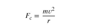
Centrifugation is based on the notion of the centrifugal force that occurs during a circular movement. Faster rotation increases sedimentation. Direct conceptual questions are prominent in entrance exams, so the students should be able to recall the order of organelle sedimentation.
Differential centrifugation is useful since it is simple and fast. However, the process does not produce entirely pure organelle fractions. Some organelles can overlap due to comparable sizes or densities. For instance, lysosomes and mitochondria may contaminate each other during separation.
This constraint has led to the creation of density gradient centrifugation, which has improved purifying accuracy.
Advantages of Density Gradient Centrifugation
Density gradient centrifugation separates organelles by buoyant density instead of only particle size. The approach employs a gradient medium such as sucrose or caesium chloride. Organelles float through the gradient until they find an area of similar density.
The approach yields highly purified cell fractions. Mitochondria, lysosomes, peroxisomes and ribosomal subunits may be separated more precisely than by differential centrifugation. This is a common method used in molecular biology labs to separate DNA, RNA and virus particles.
In cell biology, the discussion is typically on two major forms:
Rate-Zonal Centrifugation
Particles are separated by rate-zonal centrifugation largely by size and mass. A density gradient is stratified, and larger particles sediment more quickly than smaller ones. The centrifugation is interrupted before the particles fall to the bottom.
Isopycnic Centrifugation
Isopycnic centrifugation separates particles by density only. Organelles or nucleic acids migrate until they attain an equilibrium of density. After that, even if centrifuged for a long time, their position does not alter.
A trap in exams is to think that in every procedure, larger particles settle first. With density gradient centrifugation, the size of the particles is not as critical as their density. Knowing this difference will help in the better concept clarity for CUET PG 2027.
Cell Organelles Isolated by Cell Fractionation
Scientists can use cell fractionation to separate out particular organelles and analyse them individually. Each organelle sediments at a particular rate of centrifugation due to variances in size and density.
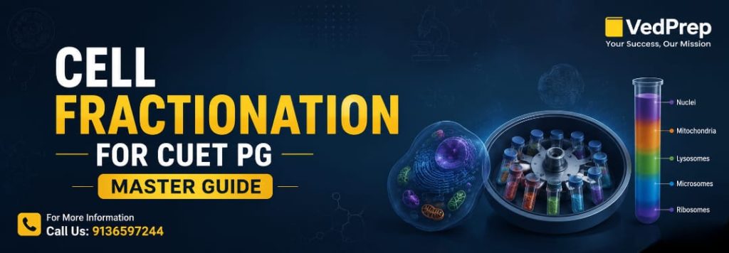
The largest and heaviest organelle is the nucleus; it is usually the first to be isolated. Mitochondria and chloroplasts sediment at intermediate rates. Lysosomes and peroxisomes demand comparatively larger centrifugal forces. Ribosomes are suspended until ultracentrifugation is administered.
Students studying for CUET PG need to carefully remember the basic order:
- Nuclei
- Mitochondria and chloroplasts
- Lysosomes and peroxisomes
- Microsomes
- Ribosomes
Microsomes are not real cell organelles. They are artificial pieces of the endoplasmic reticulum resulting from homogenization. This detail is often asked in competitive examinations.
Cell fractionation was also an important contributor to biochemical findings. Only separated mitochondrial fractions showing ATP production activity were associated with oxidative phosphorylation. Lysosomal digesting enzymes were also found in pure lysosome fractions.
These experimental links make cell fractionation an essential link between structure and function in cell biology.
Applications of Cell Fractionation in Medicine and Research
Cell fractionation is widely utilized in biological research, biotechnology, medicine and molecular diagnostics. “This technique allows scientists to study organelle-specific enzymes, proteins, nucleic acids and metabolic pathways in a controlled environment.
Mitochondrial isolation is critical in research dealing with ageing, neurological disorders, and cellular respiration. The isolation of ribosomes is useful for studies on protein synthesis and the development of antibiotics. Nuclear fractionation is a technique that scientists can use to study DNA replication and gene regulation.
In clinical laboratories, cell fractionation is used to discover biochemical abnormalities associated with lysosomal storage disorders and mitochondrial diseases. Fractionation is used in pharmaceutical companies for drug testing and the analysis of intracellular drug targets.
An example of useful research is cancer research. Researchers frequently separate nuclei and mitochondria from tumour cells to investigate aberrant metabolic activity. This technique is useful for identifying pathways underlying unregulated cell division.
Students should also know that cell fractionation is a requirement for modern molecular biology techniques such as western blotting, proteomics and organelle-specific enzyme assays. Contamination between organelles due to improper fractionation might cause misleading experimental results.
Thus, the subject is of more than theoretical interest; it is intimately related to actual biomedical research.
Cell Fractionation: Limitations and Misconceptions
Cell fractionation is very beneficial, yet the approach has key limits that trainees typically forget. Homogenization may tear organelles and lead to the leakage and contamination of enzymes. Certain cellular structures also display overlapping sedimentation features, which complicate the purification.
Often, people think that by centrifuging, you would have a pure organelle preparation. In practice, differential centrifugation often yields mixed fractions. Purity depends on proper protocol design, gradient choice and timing of centrifugation.
Another oversimplification is the notion that all cells respond similarly during fractionation. Plant, bacterial and animal cells differ in structure, and this influences the procedures used to disrupt them. Ignoring these variances decreases the accuracy of the experiment.
Another important factor is the temperature regulation. Fractionation is often done at moderate temperatures to minimize enzyme breakdown and undesired biochemical reactions. Students usually memorize the centrifugation rates and forget about temperature regulation, which is equally critical in laboratory practice.
CUET PG 2027 Conceptual comprehension over rote learning for competitive exams. More and more questions demand that students explain why a procedure works, when it fails, and how scientists make experiments more reliable.
VedPrep provides applicants with frequent concept-focused training and previous year question analysis to prepare for analytical biology themes for CUET PG, CSIR NET, IIT JAM, GATE and Assistant Professor examinations.
Cell fractionation is among the most essential elements of cell biology for CUET PG 2027 as it relates the structure of the cell to its metabolic function. A good understanding of homogenization, differential centrifugation, density gradient centrifugation and organelle isolation helps the students to answer both theory and application-based questions in competitive exams.
Frequently Asked Questions
2. Why is cell fractionation important in cell biology?
Cell fractionation helps scientists study the structure and function of individual organelles separately. By isolating mitochondria, ribosomes, or nuclei, researchers can analyze biochemical activities such as ATP production, protein synthesis, and DNA replication. The technique plays a major role in molecular biology, genetics, and medical research.
3. What is the first step in cell fractionation?
The first step in cell fractionation is homogenization. During homogenization, cells are disrupted carefully to release intracellular organelles into a solution called homogenate. Scientists use isotonic solutions during this process to prevent organelle damage caused by osmotic imbalance.
4. What is homogenate in cell fractionation?
Homogenate is the mixture obtained after cells are broken during homogenization. It contains cytoplasm, organelles, enzymes, and cellular debris suspended in a suitable medium. The homogenate is later subjected to centrifugation to separate individual organelles according to their sedimentation properties.
5. What is centrifugation in cell fractionation?
Centrifugation is the process of rotating the homogenate at high speed to separate cellular components. Larger and denser particles settle first as pellets, while smaller particles remain in the supernatant. Different centrifugation speeds help isolate specific organelles from the cell mixture.
6. What is differential centrifugation?
Differential centrifugation is a separation technique where the homogenate is centrifuged repeatedly at increasing speeds. Larger organelles like nuclei sediment first, followed by mitochondria, lysosomes, and ribosomes. The method is commonly used in cell biology laboratories and competitive exam questions.
7. What is density gradient centrifugation?
Density gradient centrifugation separates organelles according to buoyant density using gradient media such as sucrose or cesium chloride. Organelles move until they reach a region with matching density. The technique provides better purification than differential centrifugation and is widely used in advanced biological research.
8. Which organelle sediments first during centrifugation?
The nucleus sediments first during centrifugation because it is the largest and heaviest organelle in most eukaryotic cells. Low-speed centrifugation usually separates nuclei and cell debris, while smaller organelles remain suspended in the supernatant for further separation steps.
9. Why are isotonic solutions used during cell fractionation?
Isotonic solutions maintain equal osmotic pressure inside and outside organelles. This prevents swelling or shrinkage of organelles during homogenization. Sucrose solution is commonly used because it protects mitochondria, lysosomes, and other structures from osmotic damage during the fractionation process.
10. What are microsomes in cell fractionation?
Microsomes are vesicle-like membrane fragments formed from the endoplasmic reticulum during homogenization. They are not true cell organelles but appear artificially during cell disruption. Microsomes are important in biochemical studies related to detoxification enzymes and protein synthesis.
11. How are mitochondria isolated during cell fractionation?
Mitochondria are isolated through medium-speed centrifugation after nuclei have already sedimented. The mitochondrial pellet may also contain lysosomes and peroxisomes, so additional purification methods, such as density gradient centrifugation, are often required for better isolation accuracy.
12. What is the role of ultracentrifugation in cell fractionation?
Ultracentrifugation uses extremely high rotational speeds to separate very small particles such as ribosomes, viruses, and nucleic acids. It generates strong centrifugal force that allows precise isolation of low-mass cellular components which cannot sediment during ordinary centrifugation.