
The Ultimate Guide to Protein Biology in 2026: Structure, Synthesis, and Biological Significance
The biological sciences are undergoing a massive revolution in 2026. With the integration of advanced artificial intelligence and next-generation sequencing, our understanding of the cellular universe has never been more profound. At the very center of this molecular renaissance is one specific biomacromolecule that dictates the form, function, and future of every living organism.
While many textbooks offer a simplified overview of how these macromolecules are synthesized, a true mastery of the subject requires diving into the deep biochemical waters. Unlike basic summaries that merely touch upon transcription and translation, this definitive guide will explore the complete lifecycle of these molecules. We will dissect their complex chemical architecture, the intricate multi-stage process of their cellular manufacturing, the disastrous consequences of their misfolding, and the cutting-edge therapeutic applications dominating 2026.
Whether you are a postgraduate life sciences student, a competitive exam aspirant, or a curious scientific mind, this comprehensive guide will equip you with a profound understanding of life’s most versatile building blocks.
What Exactly is a Protein? The Biochemical Perspective
To truly appreciate the complexity of cellular machinery, we must first define the fundamental nature of a Protein. Biochemically, it is a highly complex, nitrogenous organic macromolecule composed of one or more long chains of amino acid residues. These chains fold into highly specific three-dimensional shapes that determine their ultimate biological function.
Amino Acids: The Fundamental Building Blocks
The alphabet of life consists of 20 standard amino acids. Each amino acid possesses a central alpha-carbon atom bonded to four distinct chemical groups: an amino group (-NH2), a carboxyl group (-COOH), a hydrogen atom, and a variable side chain known as the R-group.
The R-group is the defining feature of each amino acid, dictating its size, charge, and chemical reactivity.
- Nonpolar, Aliphatic R-groups: Such as Leucine, Isoleucine, and Valine, which tend to cluster in the interior of the folded molecule to avoid water (the hydrophobic effect).
- Aromatic R-groups: Such as Phenylalanine, Tyrosine, and Tryptophan, which possess bulky ring structures and often participate in stabilizing interactions.
- Polar, Uncharged R-groups: Such as Serine and Glutamine, which can form hydrogen bonds with water or other cellular molecules.
- Positively and Negatively Charged R-groups: Such as Lysine (positive) and Glutamate (negative), which frequently form ionic bonds or “salt bridges” crucial for structural stability.
Peptide Bonds and Polypeptide Chains
Amino acids are covalently linked together through a specialized condensation reaction. The carboxyl group of one amino acid reacts with the amino group of the next, releasing a molecule of water and forming a rigid, planar peptide bond. When dozens, hundreds, or even thousands of amino acids are linked in this continuous, unbranched sequence, they form a polypeptide chain. A single macromolecule may consist of one polypeptide chain or multiple chains interacting together.
The Four Levels of Protein Structure
Understanding how a linear string of amino acids transforms into a highly specific molecular machine is one of the most critical concepts in biochemistry. The architecture of a Protein is traditionally divided into four distinct hierarchical levels of structural organization.
Primary Structure
The primary structure refers to the exact, linear sequence of amino acids in the polypeptide chain. This sequence is directly dictated by the genetic information encoded within the DNA. Even a single alteration in this sequence—a point mutation—can have catastrophic consequences. For instance, the substitution of a single glutamic acid with valine in the beta-globin chain leads to Sickle Cell Anemia, drastically altering the molecule’s behavior.
Secondary Structure
As the polypeptide chain emerges from the ribosome, it begins to fold into localized, regular arrangements stabilized by hydrogen bonds between the backbone atoms (the carbonyl oxygen and the amide hydrogen).
- Alpha Helices: The chain coils into a right-handed spiral, with the R-groups protruding outward. This structure is highly stable and frequently found in membrane-spanning regions.
- Beta Pleated Sheets: Segments of the polypeptide chain align side-by-side, forming a sheet-like structure. These strands can run parallel or antiparallel to each other and provide immense tensile strength, as seen in silk fibroin.
Tertiary Structure
The tertiary structure is the overall three-dimensional shape of a single polypeptide chain. This is the stage where the molecule achieves its functional conformation. The folding is driven by the interactions between the diverse R-groups. The hydrophobic effect forces nonpolar side chains into the core, while polar side chains remain on the surface.
Additional stability is provided by disulfide bridges (covalent bonds between cysteine residues), electrostatic interactions, and van der Waals forces. In 2026, artificial intelligence models like AlphaFold 3 can predict these intricate tertiary structures with astonishing atomic-level accuracy.
Quaternary Structure
Many complex enzymes and structural components are not functional as single polypeptide chains. The quaternary structure refers to the spatial arrangement of multiple polypeptide subunits into a single, functional Protein complex. Hemoglobin, the oxygen-carrying molecule in our blood, is a classic example, consisting of two alpha and two beta subunits working in highly coordinated, allosteric harmony.
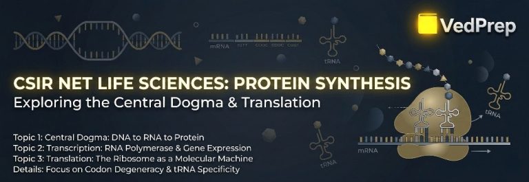
The Central Dogma: How Cells Manufacture Protein
[Image of Central Dogma of molecular biology diagram]
The synthesis of these vital macromolecules is a multi-step marvel of biological engineering, governed by the Central Dogma of Molecular Biology: DNA is transcribed into RNA, which is then translated into a polypeptide.
The Genetic Code of Protein
The instructions for building every macromolecule are stored in the sequence of nucleotides in DNA. The genetic code is read in triplets, or codons. Each sequence of three mRNA nucleotides specifies a particular amino acid. The code is degenerate (multiple codons can code for the same amino acid) and nearly universal across all domains of life, showcasing the shared evolutionary history of life on Earth.
Transcription: From DNA to mRNA
Transcription is the process of synthesizing a messenger RNA (mRNA) copy from a specific DNA template. This occurs in the nucleus of eukaryotic cells.
- Initiation: The process begins when specific transcription factors bind to a promoter region (like the TATA box) upstream of the gene. RNA Polymerase II then binds to this complex, unwinding a small section of the DNA double helix.
- Elongation: RNA Polymerase moves along the template DNA strand in a 3′ to 5′ direction, synthesizing a complementary RNA strand in the 5′ to 3′ direction. Uracil (U) is incorporated opposite Adenine (A) instead of Thymine (T).
- Termination: Once the polymerase reaches a termination signal, it detaches from the DNA, releasing the newly formed pre-mRNA transcript.
mRNA Processing: Preparing for the Cytoplasm (Protein)
In eukaryotes, the pre-mRNA must undergo extensive processing before it can serve as a template.
- 5′ Capping: A modified guanine nucleotide (7-methylguanosine) is added to the 5′ end. This protects the transcript from degradation and serves as a recognition signal for the ribosome.
- Polyadenylation: A tail of 100-250 adenine nucleotides is added to the 3′ end, further enhancing stability and regulating the mRNA’s lifespan in the cytoplasm.
- Splicing: Spliceosomes (complexes of snRNAs and specialized factors) remove the non-coding regions (introns) and join the coding regions (exons) together. Alternative splicing allows a single gene to produce multiple different polypeptide variants, vastly increasing cellular complexity.
Translation: The Assembly Line of Protein Synthesis
Once the mature mRNA enters the cytoplasm, it encounters the ribosome—the cellular factory responsible for translating the nucleotide language into the language of amino acids. Translation requires mRNA, ribosomes, and transfer RNA (tRNA) molecules that carry specific amino acids.
Amino acid Activation
Before translation can begin, tRNA molecules must be “charged” with their correct amino acids. This highly specific reaction is catalyzed by a family of enzymes called aminoacyl-tRNA synthetases, utilizing ATP to form a high-energy ester bond between the amino acid and the tRNA.
Initiation: Setting the Stage of Protein
In eukaryotes, the small ribosomal subunit, carrying the initiator methionine tRNA (recognizing the AUG start codon), binds to the 5′ cap of the mRNA. It scans along the mRNA until it locates the first AUG sequence within a favorable context (the Kozak sequence). Once the start codon is identified, the large ribosomal subunit binds, completing the initiation complex and signaling the start of Protein construction.
Elongation: Building the Chain of Protein
The ribosome contains three specific binding sites for tRNA: the A (aminoacyl) site, the P (peptidyl) site, and the E (exit) site.
- A newly charged tRNA enters the A site, its anticodon base-pairing with the corresponding mRNA codon.
- The ribosome catalyzes the formation of a peptide bond between the amino acid in the A site and the growing chain in the P site. This peptidyl transferase activity is actually catalyzed by the ribosomal RNA itself, making the ribosome a ribozyme.
- Translocation occurs as the ribosome shifts one codon down the mRNA. The empty tRNA moves to the E site and exits, the tRNA holding the growing chain moves to the P site, and the A site is left open for the next incoming tRNA. This rapid, cyclic process rapidly elongates the polypeptide.
Termination: Completing the Masterpiece
Elongation continues until a stop codon (UAA, UAG, or UGA) enters the A site. Since there are no tRNAs that correspond to stop codons, specialized release factors bind to the A site instead. These factors trigger the hydrolysis of the ester bond linking the polypeptide to the final tRNA. The newly synthesized Protein is released into the cytoplasm, and the ribosomal subunits disassemble, ready to begin another cycle.
Post-Translational Modifications: The Finishing Touches on a Protein
A newly released polypeptide chain is often not immediately functional. To become an active, mature Protein, it must undergo various Post-Translational Modifications (PTMs). These chemical alterations dynamically regulate function, cellular localization, and degradation.
Key Types of PTMs
- Phosphorylation: The addition of a phosphate group by enzymes called kinases (usually to serine, threonine, or tyrosine residues) acts as a massive biological on/off switch, regulating signal transduction pathways and enzyme activity.
- Glycosylation: The covalent attachment of carbohydrate sugar chains to the polypeptide backbone. This is crucial for cell-surface receptors, immune system recognition, and ensuring correct folding in the endoplasmic reticulum.
- Lipidation: The addition of lipid anchors, such as myristoylation or prenylation, which tether the macromolecule securely to the cellular membrane.
- Ubiquitination: The attachment of the small molecule ubiquitin serves as a “kiss of death,” targeting old or damaged molecules for destruction by the cellular recycling center known as the proteasome.
- Proteolytic Cleavage: Many enzymes and hormones are synthesized as inactive precursors (zymogens or prohormones). For example, proinsulin must be precisely cleaved by proteases to become the active hormone insulin.
The Diverse Functions of Protein in the Human Body
The sheer versatility of these macromolecules is staggering. They are not merely passive structural components; they are the active workforce executing the genetic blueprint. Almost every physiological process relies on their precise execution.
Catalysis and Metabolic Control
Enzymes are specialized biological catalysts that accelerate chemical reactions by lowering the activation energy. Without enzymes, the metabolic reactions required to sustain life would occur too slowly. DNA polymerase, amylase, and ATP synthase are all examples of enzymatic powerhouses.
Structural Support and Integrity
Structural macromolecules provide the scaffolding for cells, tissues, and entire organisms. Collagen, the most abundant macromolecule in the human body, provides immense tensile strength to skin, bones, tendons, and ligaments. Keratin forms the tough, waterproof structure of hair and nails, while the cytoskeleton (composed of actin and tubulin) maintains cellular shape and facilitates intracellular transport.
Transport and Storage
Many molecules require specialized carriers to navigate the aqueous environment of the bloodstream. Hemoglobin is a classic transport Protein that shuttles oxygen from the lungs to peripheral tissues. Lipoproteins transport hydrophobic lipids and cholesterol, while membrane channels and pumps strictly regulate the passage of ions and nutrients into and out of the cell. Ferritin serves as a specialized storage container for iron, preventing cellular toxicity.
Immunity and Defense
The immune system relies heavily on specific macromolecules to defend against invading pathogens. Antibodies (immunoglobulins) are Y-shaped molecules produced by B-cells that precisely recognize and neutralize foreign antigens, such as viruses and bacteria. Furthermore, the complement system is a complex cascade of blood plasma components that actively destroy pathogenic cells.
Communication and Signaling
Hormones such as insulin and human growth hormone function as long-distance chemical messengers, coordinating physiological responses across different organs. Furthermore, cellular receptors embedded in the plasma membrane receive these external signals and transmit the information to the cell’s interior, triggering a cascade of intracellular responses.
Protein Misfolding and Diseases
The native, functional three-dimensional shape of a polypeptide is thermodynamically delicate. Cellular stress, genetic mutations, or environmental factors can disrupt this folding process. When a molecule fails to fold correctly, it loses its biological function and often exposes sticky, hydrophobic regions that should remain buried.
Chaperones and Quality Control for Protein
To combat misfolding, cells possess a sophisticated quality control system utilizing molecular chaperones (like the Heat Shock family). These specialized cylindrical complexes provide a safe, isolated environment for nascent or unfolded chains to achieve their correct conformation without aggregating with their neighbors.
Neurodegenerative Consequences
When the quality control system is overwhelmed, the consequences are severe. Misfolded chains can aggregate into highly stable, insoluble clumps called amyloid fibrils. These toxic aggregates physically disrupt cellular architecture and function, leading to massive cell death.
- Alzheimer’s Disease: Characterized by the extracellular accumulation of Amyloid-beta plaques and intracellular neurofibrillary tangles of hyperphosphorylated tau.
- Parkinson’s Disease: Characterized by the aggregation of alpha-synuclein into Lewy bodies within dopaminergic neurons.
- Prion Diseases: Conditions like Creutzfeldt-Jakob Disease (CJD) are unique. They are caused by a misfolded Protein (the prion) that possesses the terrifying ability to induce correctly folded, normal variants to also misfold, spreading the pathology like an infectious agent without the need for DNA or RNA.
The Future of Protein Research in 2026
The landscape of biological research in 2026 is unrecognizable from a decade ago. We have transitioned from merely observing molecular biology to actively engineering it.
AI-Driven Structural Biology and Design
The release and refinement of AI models like Alpha Fold 3 and Rose TTA Fold have completely democratized structural biology. Researchers can now predict the interactions between complex subunits, DNA, RNA, and small molecule ligands with near-perfect accuracy. Furthermore, scientists are no longer restricted to the natural sequences provided by evolution. “De novo” design allows scientists to invent entirely novel, highly stable polypeptide sequences from scratch, engineered to bind specific toxins, catalyze unnatural chemical reactions, or act as next-generation biosensors.
Advanced Biologics and Targeted Degradation
The pharmaceutical industry is heavily pivoting toward biologics—therapeutic macromolecules engineered to treat previously untreatable diseases. Monoclonal antibodies have been engineered to cross the blood-brain barrier, while PROTACs (Proteolysis Targeting Chimeras) represent a revolutionary therapeutic modality. Instead of merely inhibiting a disease-causing target, PROTACs hijack the cell’s own ubiquitination machinery to permanently degrade and remove the offending Protein entirely from the cell.
Accelerate Your Academic Journey with VedPrep’s Protein Modules
Mastering the intricate details of macromolecular structure, thermodynamics, and complex synthesis pathways is an absolute necessity for anyone aspiring to crack highly competitive examinations like CSIR NET Life Sciences, GATE, or IIT JAM. Reading dense textbooks can often leave aspirants feeling overwhelmed and disconnected from the core concepts the examiners are testing.
This is precisely where VedPrep steps in to revolutionize your preparation. As India’s premier educational technology platform for higher sciences, VedPrep is dedicated to transforming complex biological concepts into intuitive, easily digestible knowledge.
Why Choose VedPrep for Your Life Sciences Preparation?
- Expert Faculty & Specialized Curriculum: At VedPrep, you aren’t just learning from generic tutors; you are mentored by top-rankers and seasoned academicians who understand the exact pulse of national-level exams. Our curriculum extensively covers unit-wise breakdowns, ensuring that high-yield topics like macromolecular biochemistry, enzyme kinetics, and molecular biology are mastered comprehensively.
- In-Depth Analytical Focus: The modern CSIR NET exam heavily favors analytical and experimental reasoning (Part C) over rote memorization. VedPrep’s interactive live classes focus heavily on interpreting experimental data, understanding structural graphs (like the Ramachandran plot), and solving complex thermodynamic numericals.
- Comprehensive Mock Tests & Real-Time Analytics: Knowledge without practice is incomplete. VedPrep provides a rigorous All-India Test Series that simulates the exact interface and difficulty of the actual examinations. Our AI-driven analytics dashboard evaluates your performance, highlighting specific weak areas in molecular biology so you can refine your strategy efficiently.
- Dedicated Mentorship & Doubt Resolution: Biological sciences require clarity. Our 24/7 doubt-clearing ecosystem ensures that whether you are stuck on a translation initiation complex or the mechanism of a specific kinase, expert help is always available. We provide psychological and strategic support to keep you motivated throughout your journey.
Don’t leave your academic aspirations to chance. Join the thousands of successful scholars who have cleared the toughest scientific hurdles with us. Enroll in VedPrep today and let us guide you toward an illustrious career in the life sciences.
Conclusion
The study of these magnificent nitrogenous macromolecules is nothing short of studying the mechanism of life itself. From the fundamental chemistry of the amino acid peptide bond to the sophisticated regulatory mechanisms of the eukaryotic ribosome, every step in the lifecycle of these molecules showcases millions of years of evolutionary refinement.
Understanding the synthesis, structure, and subsequent post-translational modification of a Protein is not merely an academic exercise; it is the key to unlocking the mysteries of human disease and pioneering the therapeutic interventions of tomorrow. As we navigate the highly advanced scientific landscape of 2026, those who command a deep understanding of molecular biology will be the ones leading the charge in research, medicine, and bioengineering. Stay curious, study rigorously, and embrace the beautiful complexity of the cellular world.
Frequently Asked Questions (FAQs)
How many standard amino acids exist in nature?
Ans: There are 20 standard amino acids that make up the vast majority of proteins in living organisms. The unique sequence and combination of these 20 amino acids determine the specific structure and function of the resulting molecule.
What is the difference between the primary and secondary structure of a protein?
Ans: The primary structure is the simple, linear sequence of amino acids coded by DNA. The secondary structure refers to localized folding patterns—like the spiral alpha-helix or the flat beta-pleated sheet—that form due to hydrogen bonding along the polypeptide backbone.
Why is the tertiary structure so important?
Ans: The tertiary structure is the final, fully folded three-dimensional shape of a single polypeptide chain. A protein must achieve this precise 3D shape to be biologically active and functional. If it loses this shape (denaturation), it loses its function.
Do all proteins have a quaternary structure?
Ans: No. Quaternary structure only exists in proteins that are made up of multiple polypeptide chains (subunits) that must assemble together to function. Hemoglobin and DNA polymerase are examples of proteins with quaternary structure.
What is the Central Dogma of Molecular Biology?
Ans: The Central Dogma describes the flow of genetic information within a biological system. It states that information flows from DNA to RNA (via transcription) and then from RNA to a functional Protein (via translation).
Where does transcription occur in a eukaryotic cell?
Ans: Transcription—the process of creating a messenger RNA (mRNA) copy from a DNA template—occurs inside the nucleus of eukaryotic cells.
What role does the ribosome play in protein synthesis?
Ans: The ribosome acts as the cellular factory during translation. It reads the sequence of codons on the mRNA transcript and coordinates with transfer RNA (tRNA) to link amino acids together in the correct order to form a polypeptide chain.
What are Post-Translational Modifications (PTMs)?
Ans: PTMs are chemical modifications made to a protein after it has been translated by the ribosome. Examples include phosphorylation (adding a phosphate group) and glycosylation (adding sugar chains), which regulate the molecule's activity, lifespan, and location within the cell.
What are molecular chaperones?
Ans: Molecular chaperones are specialized "quality control" proteins that assist newly synthesized or stress-damaged polypeptide chains in folding into their correct, functional three-dimensional shapes, preventing them from tangling or clumping together.