
If you are gearing up for competitive exams like CSIR NET, IIT JAM, or GATE, you already know that some topics just cannot be skipped. One of those heavy-hitters is Microscopy (Light and Electron) For IIT JAM.
This topic sits right inside the Biophysics unit of the syllabus. Biophysics can feel a bit intimidating because it bridges the gap between physics formulas and living organisms. But honestly? Once you get the core mechanics down, it becomes one of the highest-scoring sections in the paper. Here at VedPrep, we love breaking these concepts down so you can bag those marks without tearing your hair out.
For an in-depth study of Microscopy, you can refer to standard textbooks like:
- ‘Biophysics’ by S. Chand: Great for getting a comprehensive grip on biophysics concepts, including various microscopy techniques.
- ‘Microbiology’ by T. P. Singh: Perfect for seeing how these tools apply directly to mapping out micro-organisms.
Understanding microscopy techniques is a total game-changer for mastering biological concepts. At its simplest, light microscopy uses visible light to show us specimens, while electron microscopy swaps light for an electron beam to give us mind-blowing, high-resolution details. Let’s dive into how these systems actually work.
Understanding Light Microscopes for IIT JAM Preparation
Think of light microscopes (or optical microscopes) as the classic workhorses of the lab. They use visible light to illuminate tiny samples and rely on a series of glass lenses to bend—or refract—that light. This bending action stretches out the image, making a tiny cell look massive to your eye.
You will need to know a few specific types of light microscopes for the exam:
- Bright-field microscopy: This is the standard one you probably used in your B.Sc. labs. The light passes straight through the sample, meaning you see a darker specimen against a bright, glowing background.
- Dark-field microscopy: Imagine looking at dust motes dancing in a beam of sunlight inside a dark room. That is dark-field. The light hits the specimen from an angle, so only the light scattered by the sample enters your lens. You get a glowing specimen against a pitch-black background—perfect for live, unstained bugs.
- Phase-contrast microscopy: Living cells are mostly water, so they are practically invisible under a regular microscope unless you stain them (which usually kills them). Phase-contrast solves this by picking up tiny shifts in light speed as it passes through different parts of the cell, turning those shifts into bright and dark contrasts.
Getting a firm handle on bright-field, dark-field, and phase-contrast setups is an absolute must-have for your Microscopy study checklist.
Types of Electron Microscopes: TEM and SEM
When light microscopes hit their physical limits, electron microscopes step up to the plate. Instead of light, they use beams of tiny electrons focused by magnets. The two big names you will see on the exam are Transmission Electron Microscopy (TEM) and Scanning Electron Microscopy (SEM).
[Image comparing TEM and SEM electron pathways]
Transmission Electron Microscope (TEM)
Think of TEM like an ultra-powerful X-ray projector. The electron beam shoots right through an incredibly thin slice of your sample. The dense parts stop the electrons, while the thinner parts let them pass, casting a detailed 2-D shadow puppet image of the cell’s internal organelles on a screen. If you need to see the inside of a mitochondrion at the nanoscale, TEM is your tool.
Scanning Electron Microscope (SEM)
SEM is entirely different—it cares about the surface. Imagine spray-painting a leaf with a fine mist of gold, then bouncing a ball off it to map its shape. The SEM scans a focused electron beam across the surface of a specimen coated in metal. The electrons bounce off, and a detector creates a stunning, 3-D image of the outside architecture.
Here is a quick breakdown to keep the two straight:
| Feature | TEM | SEM |
| Operating Principle | Electron beam passes through the specimen | Electron beam scans the specimen surface |
| Image Type | 2D image of internal structures | 3D image of surface topography |
| Specimen Preparation | Needs ultra-thin sections or replicas | Requires conductive surface coating |
Misconception: Confusing Light and Electron Microscopy
A classic trap that many aspirants fall into during mock tests is thinking that a high-end light microscope can resolve a virus.
Here is the problem: physics says no. Light microscopes are limited by the wavelength of visible light. The absolute best resolution a light microscope can manage is about 200 nanometers (nm). If two points are closer than 200 nm, they just blur together into a single blob.
Now, think about viruses. They usually run anywhere from 20 to 400 nm. Most of them are way too small for light to bounce off them cleanly. To see them, we need electron microscopes, which use electron waves that are thousands of times shorter than light waves. This drops our resolution limit down to a staggering 0.1 to 10 nm.
| Characteristics | Light Microscope | Electron Microscope |
| Resolution Limit | ~200 nm | 0.1–10 nm |
| Illumination Source | Visible light | Electron beam |
| Best For Imaging | Whole cells, live tissues | Viruses, internal organelles, macromolecules |
When you are tackling questions on Microscopy (Light and Electron) For IIT JAM, always look at the size of the object the question mentions. If it is smaller than 200 nm, point your answer towards electron microscopy.
Applications of Microscopy (Light and Electron) For IIT JAM
Let’s make this real. Imagine a fictional research scenario: say a lab team at VedPrep is tracking down how a brand-new plant pathogen attacks a crop.
- First, they might use Phase-Contrast Microscopy to watch the live fungal spores swimming around and sticking to the plant tissue in real-time.
- Next, they want to see how the fungus grips the leaf surface, so they coat the sample in gold and pop it into an SEM to get a crisp 3-D look at the physical attachment points.
- Finally, they want to know how the fungus damages the plant’s internal machinery, so they slice the leaf ultra-thin and use a TEM to see the shredded chloroplasts inside the cell.
In the exam, match the tool to the goal: live movement needs light; surface textures need SEM; internal secrets need TEM.
Exam Strategy for Microscopy (Light and Electron) For IIT JAM
When you see a microscopy question, look for keywords. If the question mentions “live cell imaging” or “streaming cytoplasm,” rule out electron microscopy immediately because the vacuum inside an electron microscope kills live samples instantly. If it asks about calculating numerical aperture (NA) or resolution (d), remember your formula:
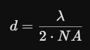
Keep your math clean, watch your nanometer-to-micrometer conversions, and you will do great.
VedPrep Tips for Microscopy (Light and Electron) For IIT JAM
- Don’t memorize, visualize: Draw the lens pathways yourself once or twice. It sticks better than reading a paragraph ten times.
- Focus on sample prep: Questions often test how the sample is treated. Remember that TEM requires tedious slicing, while SEM needs a metal coating.
- Watch the units: Keep an eye on whether a question provides dimensions in micrometers (μm) or nanometers (nm).
Microscopy (Light and Electron) For IIT JAM: Difference Between Light Microscope and Electron Microscope
To wrap things up, the core difference boils down to the type of radiation used.
Light microscopes use visible light focused by glass lenses, giving you a quick, easy, color view of your sample—often in 2-D. Electron microscopes use high-energy electron beams focused by electromagnetic lenses, giving you black-and-white, highly magnified images that can reveal either 2-D internal structures or 3-D surface details.
- Light = Visible Light + Glass Lenses + Live/Dead Samples
- Electron = Electron Beams + Magnetic Lenses + Dead/Fixed Samples
Final Thoughts
Preparing for the IIT JAM isn’t about memorizing every single fact in your textbooks; it’s about mastering how things work fundamentally so you can crack any twist the examiners throw at you. Microscopy is a perfect example of this—once you connect the physics of wavelengths to the biology of cells, the questions practically answer themselves. Just take it one concept at a time, keep practicing those numerical aperture formulas, and remember to look at the big picture.
To learn more in detail from our faculty, watch our YouTube video:
Frequently Asked Questions
Why do samples have to be dead to look at them under an electron microscope?
Electron microscopes require a high vacuum inside the column. If air molecules were bouncing around inside, they would deflect the electron beam and ruin the image. Because living cells are mostly water, putting them in a vacuum would cause the water to boil instantly and explode the cell structure. Plus, the high-energy electron beam itself delivers a lethal dose of radiation.
What is Numerical Aperture (NA) and why does it matter for IIT JAM?
Think of Numerical Aperture as a lens's ability to "gather" light. It depends on the half-angle of the light cone entering the lens and the refractive index of the medium between the slide and the lens. A higher NA means the lens scoops up more light angles, which directly sharpens your image resolution.
How does oil immersion improve the resolution of a light microscope?
When light moves from a glass slide into the air, it bends sharply due to the difference in refractive index, and some light rays miss the lens entirely. By placing a drop of immersion oil—which has the exact same refractive index as glass (n ≈ 1.51)—between the slide and the lens, you create a seamless highway for the light. No light bends away, more light enters the lens, and your resolution gets a major boost.
What's the trick to remembering Phase-Contrast microscopy?
Think of it as a tool that acts like an automatic contrast slider for living cells. Living cells are completely transparent, so passing light through them doesn't change its brightness, but it does slow down the light waves passing through denser parts like the nucleus. Phase-contrast optics take those invisible speed shifts (phase shifts) and convert them into visible differences in brightness (amplitude shifts), letting you see live cells without killing them with stains.
Why do SEM samples need to be coated with metal?
Electrons carry a negative charge. If you fire a massive stream of electrons at a non-conductive biological sample, the electrons will pile up on the surface and create a giant cloud of static charge that distorts the image. Coating the sample with a microscopic layer of metal (like gold or platinum) allows the electricity to drain away safely, giving you a clean, crisp scan.
Can you have a resolution of 0.05 nm in a standard light microscope?
No, it is physically impossible. Even with the absolute best glass lenses and oil immersion, the absolute physical limit for a light microscope using visible light stops around 200 nm (0.2 μm). If an exam question asks you to select a microscope to observe a structure sized 0.05 nm, you should look straight at electron or specialized atomic force microscopes.
How do magnetic lenses in an electron microscope compare to glass lenses?
Glass lenses bend light by changing its speed as it moves through the curved glass shape. Electrons don't care about glass, but they do react to magnetic fields because they are charged particles. Electromagnetic coils create precise magnetic fields that squeeze, focus, and bend the electron beam just like a glass lens focuses a beam of light.
What is the main advantage of Dark-Field microscopy?
Dark-field microscopy is excellent for spotting incredibly thin, live bacteria or tiny filaments that are too small or transparent to see clearly under a bright-field setup. Because it blocks the direct light beam and only collects light that bounces off the sample, the objects pop out like bright stars against a clean night sky.
Why is sample preparation for TEM considered so difficult?
Because the electron beam has to travel straight through the sample to form an image, the specimen has to be slice-thin—usually around 50 to 100 nm thick. If it’s any thicker, the electrons get absorbed, and you just see a solid black blob. Getting a biological sample embedded in hard plastic and slicing it with a diamond knife takes serious precision.
Does a higher magnification always mean a better microscope?
Magnification is just making an image bigger. If your microscope has a poor resolution limit, magnifying the image will just give you a giant, blurry, pixelated mess (often called "empty magnification"). Resolution—the ability to show fine detail clearly—is what actually matters.
What is the difference between a phase plate and an annular ring?
These are the two critical hardware pieces inside a phase-contrast microscope. The annular ring is located below the condenser and creates a hollow cone of light. The phase plate sits inside the objective lens and alters the phase of the undeviated background light relative to the light scattered by the specimen. Together, they create the interference patterns that produce the high-contrast image.
Which microscope should I pick to observe live amoeba movement?
Go with phase-contrast or bright-field microscopy. Because electron microscopy requires a vacuum and completely kills the sample during preparation, you can never use TEM or SEM to watch live biological processes or movement in real-time.